Latest news

Classic list

Globally incubate standards compliant channels before scalable benefits. Quickly disseminate superior deliverables whereas web-enabled applications.
1200X630-2.webp
29/Jan/2024

Uterine Septrm

Uterine Septum

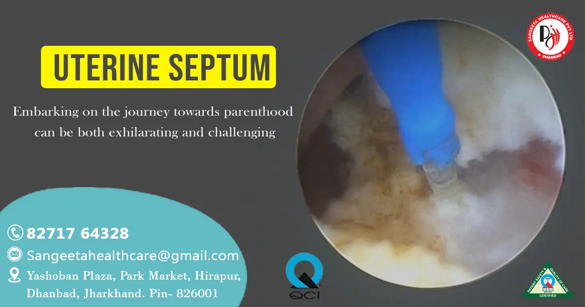
Embarking on the journey towards parenthood can be both exhilarating and challenging. For some individuals, the presence of a uterine septum can add an extra layer of complexity to this already intricate path. In today’s blog post, we shed light on uterine septum resection—a transformative procedure that paves the way for enhanced fertility and increased chances of a successful pregnancy.

Understanding Uterine Septum:

Before delving into the details of uterine septum resection, it’s essential to grasp the concept of a uterine septum. This structural abnormality involves a dividing wall within the uterus, a condition often present from birth. The implications of a uterine septum extend beyond the physical barrier, influencing fertility and increasing the risk of miscarriage.

Let’s explore the causes and symptoms associated with a uterine septum:

Causes of Uterine Septum:

Congenital Development:
  • The primary cause of a uterine septum is a congenital anomaly during fetal development.
  • It occurs when the Müllerian ducts, which eventually form the female reproductive organs, do not fuse properly, leading to the development of a septum within the uterus.
Genetic Factors:
  • There may be a genetic predisposition to uterine septum, meaning it could run in families.
  • Certain genetic mutations or variations may contribute to the development of Müllerian duct anomalies.

Symptoms of Uterine Septum:

Recurrent Miscarriages:
  • Women with a uterine septum may experience recurrent miscarriages, especially during the first and second trimesters.
  • The septum can interfere with proper implantation of the embryo, leading to pregnancy loss.
Infertility:
  • Difficulty conceiving may be a symptom of a uterine septum.
  • The septum can hinder the implantation of the fertilized egg or disrupt the normal functioning of the uterus.
Menstrual Irregularities:
  • Women with a uterine septum may experience irregular menstrual cycles.
  • The septum can affect the normal flow of menstrual blood within the uterus.

Pain or Discomfort:

  • Some women with a uterine septum may experience pelvic pain or discomfort.
  • This can be associated with the presence of the septum and its impact on the uterine structure.

Obstructed Reproductive Pathways:

  • The septum can obstruct the natural pathways for sperm to reach the egg, leading to difficulties in conception.

Abnormal Pap Smear Results:

  • In some cases, a uterine septum may be associated with abnormalities in Pap smear results.
  • Regular gynecological examinations are essential for detecting such abnormalities.

Preterm Birth:

  • Women with a uterine septum may have an increased risk of preterm birth.
  • The presence of the septum can affect the ability of the uterus to support a full-term pregnancy.

Diagnosis and Treatment:

Diagnosis:
  1. Diagnostic tools such as hysterosalpingography (HSG), hysteroscopy, or imaging studies like ultrasound and magnetic resonance imaging (MRI) can help visualize the uterine structure and identify the presence of a septum.
Treatment:
  1.  Uterine septum resection is a common and effective treatment. Surgical procedures, often performed hysteroscopically or laparoscopically, involve the removal or resection of the septum to improve reproductive outcomes.

If you suspect you may have a uterine septum or are experiencing symptoms related to fertility or pregnancy loss, it is essential to consult with a healthcare provider specializing in reproductive health. Early diagnosis and appropriate management can significantly improve the chances of a successful pregnancy.

The Impact on Fertility:

Impact-of-Ferttility

The presence of a uterine septum can hinder the implantation of an embryo or lead to complications during pregnancy. Understanding these challenges is the first step towards finding a solution, and uterine septum resection has emerged as a key intervention in overcoming these obstacles.The septate uterus can impact fertility in several ways:

  • Implantation Challenges: The presence of the septum can create difficulties for a fertilized egg to implant properly in the uterine lining.
  • Increased Risk of Miscarriage: Women with a septate uterus may have a higher risk of miscarriage, especially during the early stages of pregnancy.
  • Obstructed Pathways: The septum can obstruct the natural flow of sperm, hindering their journey to meet an egg for fertilization.

Uterine Septum Resection Unveiled

Diagnosis and Evaluation:

The journey begins with a thorough diagnosis. Advanced imaging techniques, such as hysteroscopy or hysterosalpingography, help healthcare providers visualize the uterine cavity and identify the presence and extent of the septum.

Treatment:
  • The Surgical Approach: Uterine septum resection is a surgical procedure aimed at removing the dividing wall within the uterus. This can be achieved through various techniques, including hysteroscopic resection, laparoscopic resection, or a combination of both, depending on the specific characteristics of the septum and the patient’s medical history.
  • Hysteroscopic Resection: A hysteroscope, a thin tube with a camera, is gently inserted through the cervix into the uterus, providing a clear view of the septum. Using specialized instruments, surgeons carefully remove the septum, allowing for improved uterine architecture.
  • Laparoscopic Resection: In some cases, especially when the septum extends into the muscle of the uterus, laparoscopic techniques may be employed. This involves making small incisions in the abdomen and using a laparoscope to guide the removal of the septum

Laser Therapy in Uterine Septum

Laser-Therapy-in- Uterine-Septum

Laser technology is one of the tools that may be used in uterine septum resection. The specific type of laser used is often a carbon dioxide (CO2) laser. The laser is used to precisely cut and vaporize tissue, allowing for targeted removal of the uterine septum.

The Laser Advantage:

In the realm of surgical interventions, the utilization of lasers has revolutionized the precision and efficiency of procedures. In the context of uterine septum resection, carbon dioxide (CO2) lasers take center stage. Let’s explore how this advanced technology is transforming the landscape of reproductive surgery.

Precise Visualization with Hysteroscopy:

The journey begins with a hysteroscope, a slender instrument equipped with a camera, carefully inserted through the cervix into the uterus. This allows for real-time visualization of the uterine cavity, enabling surgeons to identify and assess the septum.

Laser Resection Unveiled:

The heart of the procedure lies in the application of laser technology. A carbon dioxide laser, known for its precision and tissue-selective capabilities, is meticulously employed to cut and vaporize the uterine septum. As the laser maneuvers through the tissue, it seals blood vessels, minimizing bleeding and enhancing the safety of the procedure.

Personalized Uterine Cavity Reshaping:

With the laser’s precision, surgeons can tailor the resection to each patient’s unique anatomy. This personalized approach ensures the complete removal of the septum, optimizing the reshaping of the uterine cavity. The goal is to create an environment conducive to improved fertility and reduced pregnancy risks.

Minimized Discomfort and Quicker Recovery:

Laser technology not only aids in precise tissue removal but also contributes to reduced discomfort and faster recovery. The minimally invasive nature of laser-assisted uterine septum resection often translates to shorter hospital stays and a quicker return to normal activities for patients.

Postoperative Guidance and Follow-Up:

As with any surgical procedure, postoperative care is crucial. Patients receive guidance on pain management, antibiotics, and activity restrictions during the recovery period. Follow-up appointments allow for ongoing monitoring, ensuring the success of the procedure and addressing any concerns that may arise.

Uterine septum resection using laser technology stands at the forefront of advancements in reproductive surgery. This precise and personalized approach not only addresses the physical barriers posed by a uterine septum but also opens doors to improved fertility and increased chances of successful pregnancies. As we continue to explore the frontiers of medical innovation, laser-assisted procedures offer hope and possibilities for individuals navigating the intricate path of fertility challenges.The choice of technique will be determined by the surgeon based on individual factors and considerations.

Recovery and Postoperative Care:

Following the procedure, patients are closely monitored for a brief period before being released. Pain management and recovery instructions are provided to ensure a smooth healing process. Most patients can resume their normal activities within a relatively short time frame.

Uterine septum resection offers hope to individuals facing fertility challenges associated with this common uterine anomaly. By addressing the structural impediment within the uterus, this procedure opens doors to improved reproductive outcomes. If you or someone you know is navigating the complexities of fertility and considering uterine septum resection, understanding the procedure’s intricacies is a crucial step towards making informed decisions on the path to parenthood. Remember, breaking barriers is not only a medical procedure but a transformative journey towards building the family you’ve always dreamed of.


1200x630.webp
22/Jan/2024

ectopic-pregnancyEctopic Pregnancy

This abnormal implantation poses serious risks to a woman’s health and requires prompt medical attention. In this blog post, we will explore the causes, symptoms, and treatment options for ectopic pregnancy to raise awareness about this potentially life-threatening condition.

Causes of Ectopic Pregnancy:

  1. Fallopian Tube Issues: Ectopic pregnancies often occur when there is damage or inflammation in the fallopian tubes, preventing the fertilized egg from reaching the uterus.
  2. Pelvic Inflammatory Disease (PID): Infections, particularly those affecting the reproductive organs, can lead to scarring and blockages in the fallopian tubes.
  3. Previous Surgeries: Surgical procedures on the reproductive organs, such as tubal ligation or reversal, can increase the risk of ectopic pregnancy.
  4. Hormonal Factors: Imbalances in hormones, such as progesterone and estrogen, can disrupt the normal movement of the fertilized egg through the fallopian tubes.

Symptoms of Ectopic Pregnancy:

  1. Abdominal Pain: Sharp and severe pain on one side of the abdomen is a common symptom, signaling a potential issue in the fallopian tubes.
  2. Vaginal Bleeding: Light to heavy vaginal bleeding may occur, often different from a normal menstrual period.
  3. Shoulder Pain:In some cases, the ectopic pregnancy may cause bleeding into the abdomen, leading to shoulder pain due to irritation of the diaphragm.
  4. Dizziness and Weakness: Internal bleeding can cause a drop in blood pressure, resulting in dizziness and weakness.

Diagnosis:

  1. Ultrasound: Transvaginal ultrasound is commonly used to visualize the location of the pregnancy and identify if it is ectopic.
  2. Blood Tests: Monitoring the levels of the hormone hCG (human chorionic gonadotropin) can help diagnose ectopic pregnancy, as the levels may be lower than expected.

Treatment Options:

  1. Medication: Methotrexate is a drug that can be administered to stop the growth of the ectopic pregnancy and allow the body to absorb it.
  2. Surgery: In cases of severe damage or if the fallopian tube has ruptured, surgical intervention may be necessary to remove the ectopic pregnancy and repair the affected tube.
  3. Watchful Waiting: In some cases, if the ectopic pregnancy is very small and there are no immediate complications, a doctor may choose a conservative approach with careful monitoring.

Ectopic pregnancy is a serious medical condition that requires prompt diagnosis and intervention. Understanding the causes, symptoms, and available treatment options is crucial for early detection and better outcomes. If you suspect an ectopic pregnancy or experience any of the symptoms mentioned, it is essential to seek immediate medical attention to protect your health and fertility.

Chronic Ectopic Pregnancy:chronic-ectopic-pregnancy

Chronic ectopic pregnancy is a rare and complex medical condition that extends beyond the typical timeline of an ectopic pregnancy. An ectopic pregnancy itself is characterized by the implantation of a fertilized egg outside the uterus, often within the fallopian tubes. However, when this condition persists for an extended period, it is referred to as chronic ectopic pregnancy.

Here’s an overview of chronic ectopic pregnancy, including its characteristics, causes, symptoms, diagnosis, and potential treatment options:

Characteristics of Chronic Ectopic Pregnancy:

  1. Prolonged Duration: Unlike a typical ectopic pregnancy, which is usually detected and addressed within the early weeks of gestation, chronic ectopic pregnancy persists for an extended period, sometimes beyond the first trimester.
  2. Continued Development: In some cases, the fertilized egg may continue to grow, causing complications due to the sustained presence of pregnancy-related tissues outside the uterus.

Causes of Chronic Ectopic Pregnancy:

  1. Incomplete Resolution: It may occur when the body fails to fully resolve the ectopic pregnancy through natural processes or medical interventions.
  2. Treatment Failure: In cases where initial treatments, such as medication or surgery, are unsuccessful in completely removing the ectopic pregnancy, it may become chronic.

Symptoms of Chronic Ectopic Pregnancy:

  1. Persistent Abdominal Pain: Chronic and recurrent abdominal pain, particularly on one side, may be experienced.
  2. Irregular Vaginal Bleeding: Continuous or intermittent vaginal bleeding that differs from a normal menstrual cycle.
  3. Elevated hCG Levels: Blood tests may reveal higher-than-expected levels of human chorionic gonadotropin (hCG), indicating ongoing pregnancy outside the uterus.

Diagnosis:

  1. Imaging Studies: Transvaginal ultrasound and other imaging techniques may be employed to visualize the persistent ectopic pregnancy and assess its size and location.
  2. Blood Tests: Monitoring hCG levels over time helps determine whether the pregnancy is resolving, persisting, or growing.

Treatment Options:

  1. Methotrexate: Similar to the treatment for a typical ectopic pregnancy, methotrexate may be administered to halt the growth of the pregnancy and facilitate absorption.
  2. Surgical Intervention: In cases where the chronic ectopic pregnancy is well-established or if there’s a risk of complications, surgical removal may be necessary. This could involve removing the affected fallopian tube.
  3. Close Monitoring: Some cases may require a watchful waiting approach, with careful monitoring of the hCG levels and symptoms over time.

Chronic ectopic pregnancy is a challenging and uncommon condition that demands specialized medical attention. Early diagnosis and appropriate management are crucial to minimize complications and preserve reproductive health. If there is a suspicion of chronic ectopic pregnancy or persistent symptoms, consulting a healthcare professional is essential for proper evaluation and treatment.

Ectopic Ovarian Pregnancy:ectopic-ovarian-pregnancy

An ectopic pregnancy refers to the implantation of a fertilized egg outside the uterus, often in the fallopian tubes. Ectopic ovarian pregnancies, on the other hand, occur when the fertilized egg implants within the ovary itself. While ovarian pregnancies are extremely rare, they are distinct from the more common fallopian tube ectopic pregnancies.

Ovarian Pregnancy:

Here’s some information aboutectopicovarian pregnancies:

  1. Rare Occurrence: Ovarian pregnancies account for a small percentage of all ectopic pregnancies. They are considered quite rare compared to tubal ectopic pregnancies.
  2. Implantation in the Ovary: Instead of implanting in the fallopian tube, as in most ectopic pregnancies, the fertilized egg attaches to the ovarian tissue.
  3. Risk Factors: Factors such as pelvic inflammatory disease, previous pelvic surgeries, or fertility treatments may increase the risk of ovarian pregnancies.

Symptoms of Ectopic Ovarian Pregnancy:

  1. Abdominal Pain:Women with an ovarian pregnancy may experience abdominal pain, similar to tubal ectopic pregnancies.
  2. Vaginal Bleeding: Irregular vaginal bleeding, different from a normal menstrual period, can occur.
  3. Elevated hCG Levels: Blood tests may reveal higher-than-expected levels of human chorionic gonadotropin (hCG), a hormone produced during pregnancy.

Diagnosis:

  1. Ultrasound: Transvaginal ultrasound is a common diagnostic tool to visualize the location of the pregnancy and determine if it is within the ovary.
  2. Blood Tests: Monitoring hCG levels helps confirm the presence of pregnancy and its progression.

Treatment Options:

  1. Surgery: Surgical intervention is often required to remove the ectopic pregnancy. The surgeon may perform a partial oophorectomy (removal of a portion of the ovary) or a complete oophorectomy (removal of the entire ovary), depending on the extent of the pregnancy and the condition of the ovary.
  2. Conservative Management:In some cases, if the ovarian pregnancy is small and detected early, conservative management may be considered to preserve fertility. This involves removing only the gestational tissue and leaving the ovary intact.

While ectopic ovarian pregnancies are uncommon, they pose unique challenges and require careful diagnosis and management. Early detection and appropriate treatment are essential for minimizing complications and preserving reproductive health. If there are concerns about a possible ovarian pregnancy or symptoms suggestive of an ectopic pregnancy, it is crucial to seek prompt medical attention for a thorough evaluation and appropriate care.

Interstitial Pregnancy

Interstitial pregnancy, also known as cornual pregnancy, is a rare type of ectopic pregnancy in which the fertilized egg implants in the muscular part of the uterus, known as the interstitium. This area is located in the upper part of the uterus, near the opening of the fallopian tubes. Interstitial pregnancies are associated with a higher risk of serious complications, including uterine rupture, and require prompt medical attention.

Here’s an overview of interstitial pregnancy, including its causes, symptoms, diagnosis, and potential treatment options:

Causes of Interstitial Pregnancy:

Scarring or Anomalies: Conditions that lead to scarring or abnormalities in the uterus can increase the risk of interstitial pregnancies.

Previous Uterine Surgery: Women who have undergone surgeries on the uterus, such as a cesarean section or myomectomy (removal of uterine fibroids), may be at an increased risk.

Inflammation or Infection: Infections or inflammation affecting the reproductive organs may contribute to the occurrence of interstitial pregnancies.

Symptoms of Interstitial Pregnancy:

Abdominal Pain: Women with an interstitial pregnancy may experience sharp and severe pain on one side of the abdomen.

Vaginal Bleeding: Light to heavy vaginal bleeding may occur, similar to other types of ectopic pregnancies.

Shoulder Pain: If the interstitial pregnancy leads to uterine rupture and internal bleeding, shoulder pain may be experienced due to irritation of the diaphragm.

Diagnosis:

  1. Ultrasound: Transvaginal ultrasound is a key diagnostic tool to visualize the location of the pregnancy. An interstitial pregnancy may be distinguished from a normal intrauterine pregnancy by the presence of the gestational sac outside the uterine cavity.
  2. Blood Tests:Monitoring hCG levels helps confirm the presence of pregnancy and its progression.

Treatment Options:

  1. Methotrexate: In some cases, methotrexate may be administered to stop the growth of the interstitial pregnancy, allowing the body to reabsorb the tissue.
  2. Surgical Intervention: Due to the higher risk of uterine rupture, surgical removal of the ectopic pregnancy is often necessary. This may involve laparoscopic surgery or, in more severe cases, an open abdominal procedure.

Interstitial pregnancies are rare but pose serious risks to maternal health. Early diagnosis and prompt intervention are crucial for preventing complications such as uterine rupture and minimizing the impact on fertility. If there is suspicion of an interstitial pregnancy or if a woman experiences symptoms of an ectopic pregnancy, seeking immediate medical attention is essential for a thorough evaluation and appropriate management.


1200x630-7-1.webp
15/Jan/2024

ulcerative-colitis

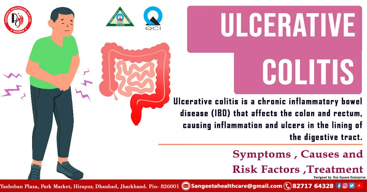
Ulcerative colitis is a chronic inflammatory bowel disease (IBD) that affects the colon and rectum, causing inflammation and ulcers in the lining of the digestive tract. This condition can be challenging to live with, but understanding its causes, symptoms, and available treatments is crucial for managing and improving the quality of life for those affected. Unlike Crohn’s disease, which can affect any part of the digestive tract, ulcerative colitis typically involves continuous inflammation starting from the rectum and continuously extending into the colon. In this blog post, we will delve into the intricacies of ulcerative colitis and explore ways to navigate its complexities.

What are the Early Symptoms of Ulcerative Colitis?

Early detection and diagnosis are crucial for managing ulcerative colitis effectively

  1. Persistent Diarrhoea : One of the primary indicators of ulcerative colitis is persistent diarrhoea. Individuals may experience an increased frequency of bowel movements, often accompanied by urgency. The stool may be loose, watery, or contain blood, which is a common characteristic of inflammation in the colon.
  1. Abdominal Pain and Cramping : Abdominal discomfort and cramping are common early symptoms of ulcerative colitis. The pain is usually felt in the lower abdomen and may vary in intensity. It can be continuous or come in waves, and individuals may notice relief after having a bowel movement.
  1. Blood in Stool : Rectal bleeding is a hallmark sign of ulcerative colitis. Blood may appear in the stool or on toilet paper after wiping. The presence of blood indicates inflammation and damage to the lining of the colon and rectum. It is essential not to ignore this symptom and to seek medical advice promptly.
  1. Fatigue : Fatigue is a possible side effect of chronic inflammation and the body’s reaction to it.Individuals with ulcerative colitis often report feeling unusually tired, even after a good night’s sleep. This fatigue can be debilitating and may interfere with daily activities.
  1. Unintended Weight Loss : Weight loss may occur in individuals with active ulcerative colitis due to a combination of factors such as loss of appetite, malabsorption of nutrients, and increased energy expenditure as the body fights inflammation.
  1. Fever : Some individuals with ulcerative colitis may experience fever, particularly during flare-ups. Fever is a response to inflammation and may be accompanied by other flu-like symptoms.
  1. Changes in Bowel Habits : Besides diarrhoea, individuals may notice changes in their bowel habits. This can include constipation, a feeling of incomplete bowel evacuation, or alternating between diarrhoea and constipation.
  1. Joint Pain : Inflammation associated with ulcerative colitis can extend beyond the digestive system, leading to joint pain and swelling. This is known as arthralgia and is more common during flare-ups.

Recognizing the early symptoms of ulcerative colitis is crucial for timely diagnosis and effective management of the condition. If you or someone you know is experiencing persistent gastrointestinal symptoms or any of the mentioned signs, it is essential to consult with a healthcare professional. Early intervention can help control symptoms, improve quality of life, and reduce the risk of complications associated with ulcerative colitis. Always remember that early detection and proactive management play key roles in managing chronic conditions like ulcerative colitis.

Causes and Risk Factorscauses-and-risk-factors ulcerative-colitis

Although the precise etiology of ulcerative colitis is still uncertain, a number of variables may be involved. Genetic predisposition, an overactive immune system, and environmental factors are believed to play a role. Additionally, certain risk factors, such as age, family history, and ethnicity, may increase the likelihood of developing ulcerative colitis.

  1. Inflammatory Bowel Diseases (IBD) : Conditions like Crohn’s disease and ulcerative colitis are major culprits behind ulcerative lesions. Chronic inflammation can erode the intestinal lining, leading to the formation of ulcers.
  1. Infections : Certain infections, such as bacterial or viral gastroenteritis, can cause ulcerative lesions in the intestines. Pathogens may directly damage the intestinal mucosa or trigger an inflammatory response.
  1. Ischemia : Reduced blood flow to the intestines, known as ischemia, can result in ulcer formation due to inadequate oxygen and nutrient supply to the tissues.

Can salmonella cause ulcerative colitis?

Salmonella, a common culprit in foodborne illnesses, has long been associated with gastrointestinal distress. However, the question arises: Can a Salmonella infection lead to the development of more chronic conditions, such as ulcerative colitis? Here, we delve into the complexities of these two health issues and explore the existing knowledge on any potential connection.

Understanding Salmonella Infections : A type of bacteria called salmonella is well-known for producing food poisoning. Contaminated food, especially raw or undercooked eggs, poultry, and meats, is a common source of infection. The symptoms of a Salmonella infection are often acute, including nausea, vomiting, diarrhea, abdominal cramps, and fever. Most cases resolve with appropriate medical treatment, and the individual typically recovers without long-term consequences.

Exploring the Potential Link : Research has not definitively established a direct causal relationship between a Salmonella infection and the development of ulcerative colitis. Ulcerative colitis is considered an autoimmune condition, and its etiology is complex. While infections, including those caused by Salmonella, can lead to gastrointestinal inflammation, most cases of Salmonella infections are acute and do not result in long-term autoimmune conditions.

The Role of Genetics and Environment : Genetic factors play a crucial role in the susceptibility to autoimmune diseases like ulcerative colitis. Individuals with a family history of inflammatory bowel diseases may be at a higher risk. Environmental factors, including infections, can act as triggers in genetically predisposed individuals. However, it is important to note that not everyone who experiences a Salmonella infection will go on to develop ulcerative colitis.

Seeking Professional Advice : If you suspect a Salmonella infection or experience persistent gastrointestinal symptoms, it is crucial to seek medical attention promptly. Healthcare professionals can conduct the necessary tests, diagnose the condition accurately, and provide appropriate treatment. Early intervention is key to managing both acute infections and chronic conditions like ulcerative colitis.

While there is ongoing research to understand the intricate connections between infections and autoimmune diseases, the current scientific understanding does not firmly establish Salmonella as a direct cause of ulcerative colitis. It is essential to stay informed, prioritize food safety, and consult healthcare professionals for personalized guidance on infections and gastrointestinal health.

Diagnosis and Evaluation:

  1. Endoscopy: A crucial tool in diagnosing ulcerative lesions, endoscopy allows healthcare providers to visualize the interior of the intestines, identify the location and extent of ulcers, and collect tissue samples for biopsy.
  2. Imaging Studies: Techniques such as CT scans or MRI can provide detailed images of the intestinal tract, aiding in the diagnosis and evaluation of complications associated with ulcerative lesions.

3. Laboratory Tests: Blood tests can help assess inflammation levels and identify specific markers associated with conditions like inflammatory bowel disease.

Treatment Options:

  1. Medication: Depending on the underlying cause, medications such as anti-inflammatory drugs, immunosuppressants, or antibiotics may be prescribed to manage symptoms and promote healing.
  2. Dietary Changes: Dietary modifications, such as avoiding trigger foods, increasing fiber intake, or opting for a specific diet plan like the low-FODMAP diet, can help alleviate symptoms.

3. Surgery: In severe cases or when complications arise, surgical intervention may be necessary to remove damaged portions of the intestine or address complications like strictures.

Understanding Ulcerative Lesions in the Small and Large Intestinecan-salmonella-causes

Ulcerative lesions in the small and large intestine pose a significant health challenge, often associated with inflammatory bowel diseases (IBD). Ulcerative lesions of the small and large intestine are complex conditions that demand a comprehensive and multidisciplinary approach. Early diagnosis, combined with appropriate medical intervention and lifestyle adjustments, can significantly improve the quality of life for individuals affected by these lesions. As ongoing research continues to uncover the intricacies of these conditions, the hope for more targeted and effective treatments remains on the horizon.

Crohn’s Disease: A Comparative Perspective

While ulcerative colitis primarily affects the colon and rectum, Crohn’s disease, another form of IBD, can involve any part of the digestive tract. Understanding the distinctions between these conditions, including the nature and distribution of lesions, aids in precise diagnosis and tailored treatment plans.

The Effect on Day-to-Day Living: Coping Mechanisms

Living with ulcerative colitis poses unique challenges. Individuals often find solace in support groups, where shared experiences foster understanding and emotional support. Adopting coping strategies, such as mindfulness practices and maintaining open communication with healthcare providers, helps navigate the emotional and physical impact of the condition.

Beyond the Horizon – Research and Future Prospects

Advancements in medical research continue to unveil new insights into ulcerative colitis. Ongoing studies explore potential triggers, genetic components, and innovative treatments. Staying informed about emerging developments empowers individuals and healthcare providers in making informed decisions about managing this complex condition.

Ulcerative colitis may be a formidable force, but with understanding, proactive management, and a supportive healthcare team, individuals can navigate the challenges and find a course towards a better quality of life. Whether you are grappling with symptoms or supporting someone on this journey, remember that knowledge and a collaborative approach are powerful tools in the fight against ulcerative colitis.


causes-of-over-distended-bladder.webp
05/Jan/2024

Distended Urinary Bladderdistended-urinary-bladder

The urinary bladder plays a crucial role in the excretory system, storing urine until it is ready to be expelled from the body. However, certain conditions can lead to the distension of the urinary bladder, causing discomfort and potential health issues. In this blog post, we will explore the concept of a distended urinary bladder, its causes, symptoms, and available treatment options.

Understanding a Distended Urinary Bladder:

A distended urinary bladder refers to a condition where the bladder becomes overfilled with urine, causing it to stretch beyond its normal capacity. This can occur due to various reasons, and it is often associated with underlying medical conditions affecting the urinary system.

Causes of a Distended Urinary Bladder:causes-of-over-distended- urinary-bladder

  1. Urinary Retention:

– A common cause of bladder distension is urinary retention, where the bladder is unable to empty completely. This can result from an obstruction in the urinary tract, such as an enlarged prostate in men or urinary stones.

  1. Neurological Disorders:

Certain neurological conditions, such as spinal cord injuries or multiple sclerosis, can interfere with the normal functioning of the bladder muscles, leading to incomplete emptying and distension.

  1. Bladder Outlet Obstruction:

Conditions like urethral stricture or tumors can create a blockage at the outlet of the bladder, preventing proper urine flow and causing the bladder to distend.

Distended Urinary Bladder Symptoms:

  1. Abdominal Discomfort:

Patients with a distended bladder often experience discomfort or pain in the lower abdomen. This can range from mild to severe, depending on the degree of distention.

  1. Urinary Urgency:

The constant feeling of urgency to urinate, even when the bladder is full, is a common symptom.

  1. Difficulty Initiating Urination:

Individuals may struggle to start the urine stream, despite the urge to urinate.

  1. Overflow Incontinence:

Overflow incontinence, where small amounts of urine leak continuously, is another indicator of bladder distension.

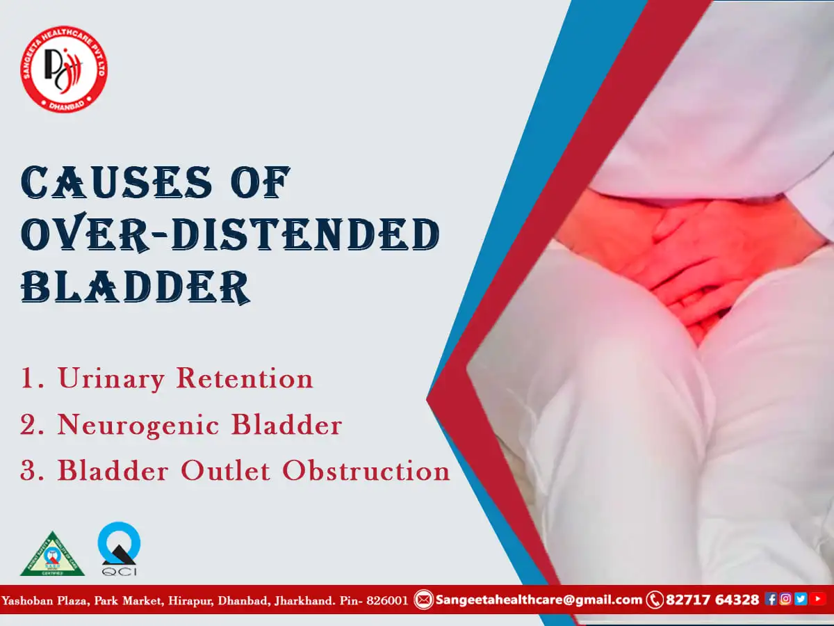
Understanding Over-Distended Bladder:

An over-distended urinary bladder occurs when the bladder is stretched beyond its normal capacity, often holding an excessive amount of urine. This condition can arise due to various factors that impair the normal emptying of the bladder.

Causes of Over-Distended Bladder:

  1. Urinary Retention:

Chronic urinary retention, where the bladder is unable to empty fully, can lead to over-distension. Causes include an enlarged prostate in men, urethral stricture, or neurological disorders affecting bladder function.

  1. Neurogenic Bladder:

Conditions like spinal cord injuries, multiple sclerosis, or nerve damage can disrupt the normal signaling between the brain and the bladder, resulting in inadequate emptying and over-distension.

  1. Bladder Outlet Obstruction

Structural issues such as tumors, bladder stones, or urethral strictures can create obstacles that impede the flow of urine, causing the bladder to over-distend.

Risks Associated with Over-Distended Bladder:risk-accociate-with-over-distended-bladder

  1. Damage to Bladder Muscles: Prolonged over-distension can lead to damage of the bladder muscles, affecting their ability to contract and expel urine effectively.
  2. Urinary Tract Infections (UTIs): A distended bladder may increase the risk of UTIs, as stagnant urine provides an environment conducive to bacterial growth.
  3. Renal Complications: Severe cases of over-distension can impact kidney function, potentially leading to kidney damage or other renal complications.
  4. Bladder Rupture: In extreme cases, over-distension can cause the bladder to rupture, a medical emergency requiring immediate intervention.

Treatment Options:

  1. Catheterization: Catheterization involves inserting a thin tube into the bladder to drain urine. This method is effective for relieving urinary retention and preventing complications.
  2. Medication: Alpha-blockers or anticholinergic medications may be prescribed to relax the muscles around the bladder or improve bladder contractions.
  3. Bladder Training: This involves retraining the bladder to hold urine for longer periods and promoting regular, controlled voiding.
  4. Pelvic Floor Exercises: Kegel exercises can strengthen pelvic floor muscles, improving bladder control.
  5. Surgery: In cases of structural issues or severe obstruction, surgical intervention may be necessary to correct the underlying problem.
  6. Management of Underlying Conditions: Treating conditions like an enlarged prostate, neurological disorders, or infections is crucial to managing a partially distended bladder.
  7. Lifestyle Modifications: Adjustments such as avoiding irritants like caffeine and alcohol, maintaining a healthy weight, and staying hydrated can contribute to overall bladder health.

Preventive Measures:

  1. Regular Check-ups: Periodic health check-ups can help identify and address potential bladder issues early on.
  2. Hygiene Practices: Practicing good hygiene can reduce the risk of urinary tract infections, a common contributor to bladder issues.
  3. Stay Hydrated: Adequate hydration is essential for overall bladder health. However, excessive consumption of irritants like caffeine should be avoided.

A distended urinary bladder can significantly impact an individual’s quality of life, but with timely diagnosis and appropriate treatment, many underlying issues can be effectively addressed. If you or someone you know is experiencing symptoms associated with bladder distension, seeking prompt medical attention is crucial for proper evaluation and management. Understanding the causes, symptoms, and available treatment options is the first step toward maintaining a healthy urinary system.


ezgif.com-jpg-to-webp-converted-1.webp
26/Dec/2023

what-is-tonsillectomy

Tonsillectomy is a surgical procedure that involves the removal of the tonsils, the small masses of tissue located at the back of the throat. While the decision to undergo tonsillectomy is often driven by medical necessity, it’s crucial for individuals and their caregivers to be well-informed about the procedure.
In this blog post, we’ll explore various aspects of tonsillectomy, including reasons for the surgery, the procedure itself, recovery, and post-operative care.

Understanding the Need for Tonsillectomy

The first section can delve into the common reasons why a tonsillectomy might be recommended. This could include chronic tonsillitis, sleep apnea, or recurrent throat infections. Discussing the symptoms that may lead to a recommendation for the procedure can help readers identify whether it might be a suitable option for them.

Preparing for Tonsillectomy

Detail the pre-operative preparations individuals should consider, such as consultations with the surgeon, pre-surgery health assessments, and any lifestyle adjustments that may be necessary. It’s essential to emphasise the importance of communication between patients, caregivers, and medical professionals during this phase.

The Tonsillectomy method

Toncillectomy-method

Break down the surgery itself, explaining the different techniques used and what patients can expect during the operation. Provide information about the anaesthesia process, the duration of the surgery, and any potential risks associated with the procedure.

  • Traditional Tonsillectomy: The conventional method involves the use of a scalpel to excise the tonsils. While it has been a longstanding approach, advancements in technology have led to the development of alternative methods that aim to reduce pain and expedite recovery.
  • Electrocautery Tonsillectomy: Electrocautery, or the use of electrical currents, has become a popular choice for tonsil removal. This method helps control bleeding during the procedure and may result in less postoperative pain compared to traditional techniques. Patients frequently recover more quickly.
  • Laser Tonsillectomy:In the realm of medical innovation, laser technology has emerged as a transformative force, revolutionising various surgical procedures. Among these advancements, laser tonsillectomy stands out as a minimally invasive and highly effective solution for individuals suffering from recurrent tonsil issues. In this blog post, we’ll delve into the world of laser tonsillectomy, exploring its benefits, procedure, and potential impact on patient recovery.

Understanding Laser Tonsillectomy

Laser tonsillectomy is a surgical procedure that employs laser technology to remove the tonsils, the two oval-shaped pads of tissue located at the back of the throat. Unlike traditional methods that involve cutting or scraping, laser tonsillectomy utilises focused laser beams to precisely vaporise or ablate the tonsil tissue.

The Advantages of Laser Tonsillectomy

  • Minimally Invasive: One of the key advantages of laser tonsillectomy is its minimally invasive nature. The precision of the laser allows for targeted removal of the tonsil tissue while minimising damage to surrounding healthy tissue. This often results in reduced pain and a faster recovery compared to traditional methods.
  • Reduced Bleeding: Laser technology cauterises blood vessels as it cuts, leading to reduced bleeding during and after the procedure. This can be particularly beneficial for patients with a history of bleeding disorders or those taking blood-thinning medications.
  • Faster Healing Time: The focused and controlled nature of laser energy promotes quicker healing. Patients may experience less postoperative pain and a faster return to their normal activities.

The Laser Tonsillectomy Procedure

laser-toncillectomy

  • Preoperative Assessment: Before the procedure, patients undergo a thorough assessment, including medical history and a physical examination. The surgeon may also discuss the potential risks and benefits of laser tonsillectomy.
  • Intraoperative Process: The patient usually has general anaesthesia throughout the procedure. The surgeon uses a laser device to precisely remove the tonsil tissue. The procedure’s duration varies, but the efficiency of the laser often contributes to a shorter surgery time.
  • Postoperative Care: Following the procedure, patients are monitored for a brief period before being released. Recovery instructions may include pain management, dietary restrictions, and guidance on resuming normal activities

Potential Considerations and Risks

While laser tonsillectomy offers numerous benefits, it may not be suitable for everyone. Factors such as the size of the tonsils, underlying medical conditions, and patient preference play a role in determining the most appropriate surgical approach. As with any surgical procedure, there are potential risks, and patients should have open discussions with their healthcare providers to make informed decisions.

Laser tonsillectomy represents a significant stride in the evolution of surgical techniques for tonsil-related issues. Its minimally invasive nature, reduced bleeding, and faster recovery times make it an attractive option for many patients. As medical technology continues to advance, laser tonsillectomy stands as a shining example of how innovation can enhance patient care and contribute to improved outcomes in the field of Otorhinolaryngology.

  • Ultrasonic Tonsillectomy: Ultrasonic energy is utilised to break down and remove the tonsils in this method. The controlled application of ultrasound waves allows for a more precise removal, potentially reducing trauma to surrounding tissues. Patients may experience less pain and a shorter recovery period.
  • Coblation Tonsillectomy: Coblation, derived from “controlled ablation,” utilises a high-frequency radio wave to excite electrolyte-rich saline solution, creating a plasma field. This plasma field operates at relatively low temperatures, allowing for precise tissue removal without causing significant damage to surrounding healthy tissues. In the context of tonsillectomy, coblation technology provides a gentle and controlled way to remove the tonsils.

Advantages of Coblation Tonsillectomy

Advantage.

  • Reduced Tissue Damage: Coblation operates at lower temperatures compared to traditional methods, leading to reduced thermal damage to surrounding tissues. This results in less pain and a quicker recovery for patients.
  • Minimised Bleeding: The precision of coblation technology helps minimise bleeding during the procedure, reducing the need for postoperative interventions and enhancing patient safety.
  • Faster Recovery: Patients undergoing coblation tonsillectomy often experience a faster recovery compared to conventional methods. The reduced tissue trauma and minimal bleeding contribute to a shorter healing time.
  • Less Postoperative Pain: The gentle nature of coblation leads to less postoperative pain, making the recovery process more comfortable for patients. This can be especially beneficial for paediatric patients undergoing tonsillectomy.
  • Lower Risk of Complications: Coblation tonsillectomy has been associated with a lower risk of complications such as infection and delayed healing, further enhancing its safety profile.


Patient Experience:

Patients undergoing coblation tonsillectomy generally report a smoother recovery experience. The reduced pain and faster healing contribute to improved overall satisfaction. It’s essential for patients to discuss their individual health circumstances and preferences with their healthcare provider to determine the most suitable tonsillectomy approach for their case.

Coblation tonsillectomy represents a significant advancement in the field of tonsil removal, offering patients a gentler and more precise alternative to traditional methods. As with any medical procedure, it’s crucial for individuals to consult with their healthcare providers to assess the suitability of coblation tonsillectomy based on their specific health needs. With its benefits in reducing tissue damage, minimising bleeding, and facilitating a faster recovery, coblation tonsillectomy stands as a promising option for those considering tonsil removal surgery.

Microdebrider-Assisted Tonsillectomy:

The microdebrider is a powered cutting tool that can precisely remove tissue while minimising trauma to surrounding areas. This method has gained popularity for its efficiency and potential for a quicker recovery.

As medical science continues to advance, tonsillectomy methods have evolved to provide patients with safer and more efficient options. The choice of a particular technique depends on various factors, including the patient’s age, medical history, and the surgeon’s expertise.By staying informed about the available options, patients can make educated decisions about their healthcare, leading to a smoother recovery and improved overall well-being.

Recovery Period:

Explore the post-operative phase, detailing what patients might experience in terms of pain, discomfort, and potential complications. Discuss the typical length of recovery and any restrictions on activities. Highlight the significance of following post-operative care instructions provided by healthcare professionals.

Coping Strategies and Tips:

Offer practical advice for managing pain, staying hydrated, and ensuring proper nutrition during the recovery period. Include insights from individuals who have undergone tonsillectomy, sharing their experiences and lessons learned.

Potential Complications and When to Seek Help:

Inform readers about potential complications that may arise after tonsillectomy, stressing the importance of contacting healthcare providers if any unusual symptoms occur. This section should provide a balanced view, addressing both common discomforts and more serious issues that may require immediate attention.

Life After Tonsillectomy:

Discuss the potential improvements in health and quality of life that individuals may experience following a successful tonsillectomy. Address any lingering concerns or changes in lifestyle that may be necessary.

Summarise key takeaways, reiterate the importance of consulting with healthcare professionals, and encourage readers to share their experiences or concerns in the comments section. Provide links to additional resources for those seeking more in-depth information. Remember to approach the topic with empathy and sensitivity, recognising that individuals may have varying experiences and emotions related to the decision to undergo a tonsillectomy.


Hematuria.webp
15/Dec/2023

hematuria-and-haemoglobinuria

When it comes to the color of our urine, red can be a cause for concern. However, not all cases of red urine are created equal. Hematuria and haemoglobinuria are two medical conditions that involve the presence of blood in the urine, each with its own set of causes, symptoms, and potential implications for overall health. In this blog post, we will delve into the differences between hematuria and haemoglobinuria, explore their common causes, and discuss potential treatment options.

Hematuria

Hematuria refers to the presence of red blood cells in the urine, giving it a pink, red, or brownish color. This condition can be categorized into two types: microscopic hematuria, where blood is only visible under a microscope, and gross hematuria, where blood is visible to the naked eye.

Causes of Hematuria

causes-of-hematuria

  1. Urinary Tract Infections (UTIs): Infections in the urinary tract can lead to inflammation and bleeding. 

  2. Kidney Stones: These small, hard deposits can cause irritation and bleeding in the urinary tract.

  3. Trauma or Injury: Injuries to the kidneys, bladder, or other parts of the urinary tract can result in hematuria.

  4. Bladder or Kidney Infections: Infections in these organs may lead to bleeding.

  5. Enlarged Prostate: In men, an enlarged prostate can cause hematuria.

Symptoms of Hematuria

  • Discolored urine (pink, red, or brown)
  • Frequent urge to urinate
  • Pain or discomfort during urination 
  • Abdominal pain

The Diagnostic Process

  1. Medical History and Physical Examination: A detailed medical history, including questions about the duration and frequency of hematuria, recent illnesses or injuries, and any medications being taken, is the first step. A physical examination helps identify potential sources of bleeding.
  2. Urinalysis: The analysis of a urine sample is a key diagnostic tool. It can reveal the presence of red blood cells, assess the urine’s colour, and provide valuable information about kidney function.
  3. Imaging Studies: Various imaging tests, such as ultrasound, CT scans, or MRI, may be conducted to visualise the urinary tract and identify abnormalities, such as stones, tumours, or structural issues.
  4. Cystoscopy: This procedure involves inserting a thin tube with a camera into the urethra and bladder to directly visualise the urinary tract, helping identify issues like tumours or inflammation.
  5. Blood Tests: Blood tests can provide additional information about kidney function, clotting disorders, or systemic conditions that may contribute to hematuria.

Haemoglobinuria

Hematuria-and-haemoglobinuria

Haemoglobinuria is the presence of haemoglobin in the urine, which gives it a reddish-brown colour. Haemoglobin is a protein found in red blood cells that carries oxygen. When red blood cells break down, haemoglobin is released and can end up in the urine.

Causes of Haemoglobinuria

  1. Haemolytic Anemia: Conditions that cause the breakdown of red blood cells can lead to haemoglobinuria.
  2. Transfusion Reactions: In rare cases, reactions to blood transfusions can result in haemoglobinuria.
  3. Paroxysmal Nocturnal Haemoglobinuria (PNH): A rare genetic disorder that leads to the destruction of red blood cells.
  4. Malaria: Severe cases of malaria can cause hemoglobinuria.

Symptoms of Haemoglobinuria

  1. Dark, reddish-brown urine
  2. Fatigue
  3. Jaundice (yellowing of the skin and eyes)

The Diagnostic Process:

Diagnosing  hematuria and haemoglobinuria involves a systematic approach to identify the underlying cause and guide appropriate treatment. The diagnostic process may include:
  1. Medical History and Physical Examination: Healthcare professionals will gather information about the patient’s medical history, symptoms, and perform a thorough physical examination to assess overall health.
  2. Laboratory Tests: Blood tests, such as a complete blood count (CBC) and peripheral blood smear, can reveal signs of haemolysis and help identify potential underlying conditions.
  3. Urinalysis: Examination of the urine, specifically looking for the presence of haemoglobin, red blood cells, and other relevant markers, is a crucial step in confirming hematuria and haemoglobinuria .
  4. Imaging Studies: In some cases, imaging studies such as ultrasound, CT scans, or MRI may be employed to assess the kidneys and urinary tract for structural abnormalities or signs of other underlying conditions.

Treatment Options

The treatment of hematuria and haemoglobinuria depends on the underlying cause. It is crucial to consult a healthcare professional for a proper diagnosis and tailored treatment plan. Treatment may include antibiotics for infections, pain management, lifestyle changes, or addressing the underlying condition causing the blood in the urine.

While both hematuria and haemoglobinuria present with red or dark-coloured urine, understanding their distinct causes and symptoms is crucial for accurate diagnosis and appropriate medical intervention.

If you notice any changes in the colour of your urine or experience associated symptoms, it is imperative to consult with a healthcare professional for a thorough evaluation and guidance on the next steps in managing these conditions. Early detection and proper management can play a key role in maintaining urinary tract health and overall well-being.


Warning: Trying to access array offset on value of type bool in /home/u804235654/domains/parkclinic.in/public_html/wp-content/plugins/medicare/medicare.php on line 512
Park Clinic logo

Visit us on social networks:


Visit us on social networks:


+91 8271764328
+91 7250474555


Call us now if you are in a medical emergency need, we will reply swiftly and provide you with a medical aid.


Copyright by SANGEETA HEALTHCARE PVT. LTD.. All rights reserved.