Latest news

Classic list

Globally incubate standards compliant channels before scalable benefits. Quickly disseminate superior deliverables whereas web-enabled applications.
1200x630-1-2-1.webp
22/Apr/2024

PCOS

pcod and pcos

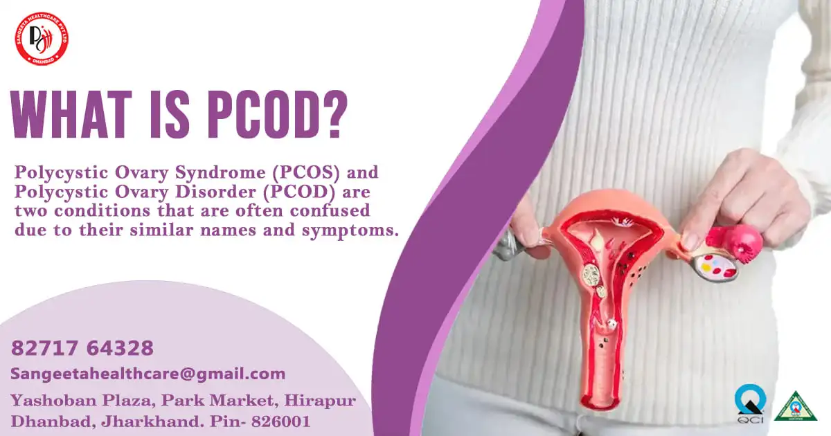
Polycystic Ovary Syndrome (PCOS) and Polycystic Ovary Disorder (PCOD) are two conditions that are often confused due to their similar names and symptoms. However, they are distinct medical conditions with their own set of characteristics and implications. In this blog post, we will discuss thoroughly about both PCOD and PCOS to shed light on these often-misunderstood conditions.

What is PCOD?

Polycystic Ovary Disorder (PCOD), also known as Polycystic Ovarian Syndrome (PCOS), is a hormonal disorder characterized by the presence of multiple small cysts on the ovaries. However, not all individuals diagnosed with PCOD necessarily have cysts. PCOD is primarily a reproductive disorder, often resulting in irregular menstrual cycles and difficulties in conceiving.

Key Features of PCOD:
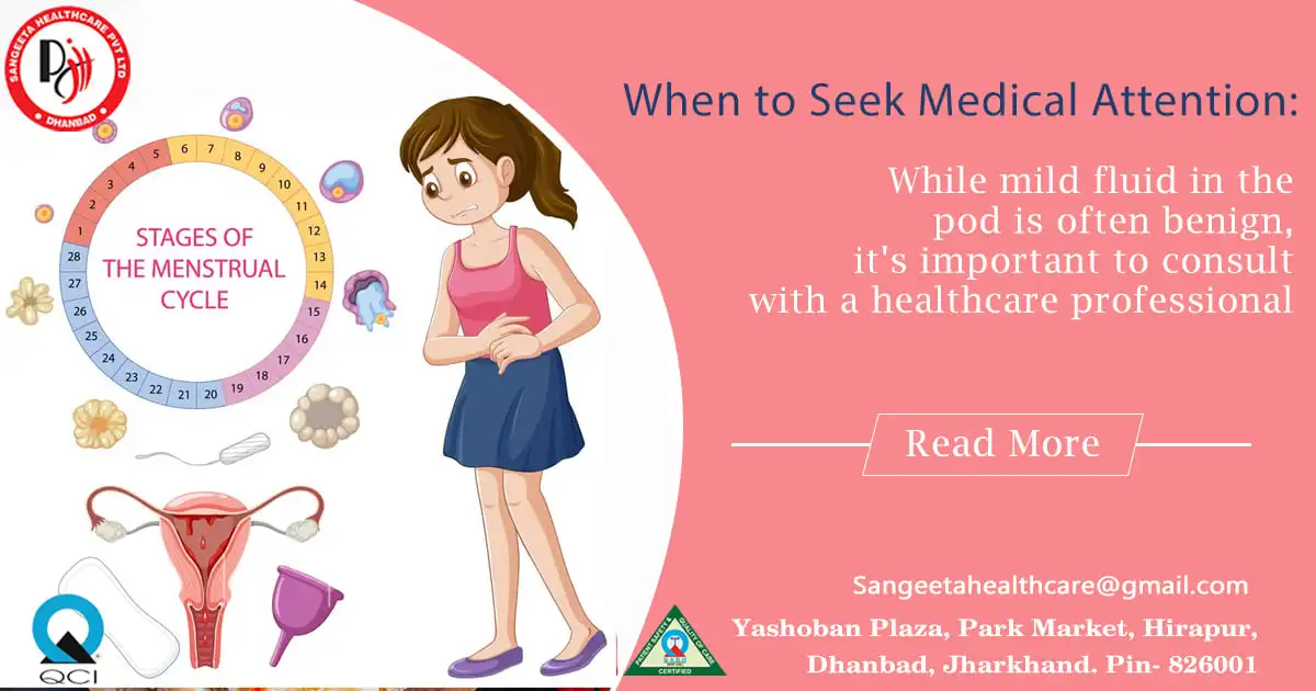
  1. Menstrual Irregularities: Women with PCOD frequently experience irregular menstrual cycles, including prolonged periods, scanty bleeding, or even absence of menstruation.
  2. Hormonal Imbalance: PCOD is associated with hormonal imbalances, particularly elevated levels of androgens (male hormones) such as testosterone.
  3. Ovarian Cysts: While the name suggests the presence of cysts, not all individuals with PCOD develop ovarian cysts. These cysts are tiny, fluid-filled pouches that grow on the ovaries.

Metabolic Issues: PCOD is often linked with metabolic disturbances such as insulin resistance, which can lead to weight gain, type 2 diabetes, and other health complications

What is PCOS?

Polycystic Ovary Syndrome (PCOS) is a broader condition that encompasses not only ovarian cysts but also a range of other symptoms related to hormonal imbalance. One of the most prevalent endocrine conditions affecting women who are fertile is PCOS.

Key Features of PCOS:
  1. Menstrual Irregularities: Similar to PCOD, women with PCOS may experience irregular menstrual cycles, often characterized by oligo menorrhea (infrequent periods) or amenorrhea (absence of menstruation).
  2. Hormonal Imbalance: PCOS is characterized by high levels of androgens, insulin resistance, and abnormalities in other hormones such as luteinizing hormone (LH) and follicle-stimulating hormone (FSH).
  3. Ovarian Cysts: The presence of ovarian cysts is common in PCOS, but they are not a prerequisite for diagnosis. These cysts are typically small and numerous, giving the ovaries a characteristic “polycystic” appearance on ultrasound.
  4. Metabolic Issues: Insulin resistance and metabolic syndrome are common in individuals with PCOS, increasing the risk of obesity, type 2 diabetes, cardiovascular disease, and other metabolic complications.

Is PCOD and PCOS same?

pcod and pcos are same

PCOD (Polycystic Ovary Disorder) and PCOS (Polycystic Ovary Syndrome) are related conditions, but they are not exactly the same. They share some similarities but also have differences that set them apart. Here’s a breakdown:

Similarities:
  1. Ovarian Cysts: Both PCOD and PCOS involve the presence of ovarian cysts, although they may vary in number and size.
  2. Hormonal Imbalance: Both conditions are characterized by hormonal imbalances, particularly elevated levels of androgens (male hormones) such as testosterone, and irregularities in other hormones like LH and FSH.
  3. Menstrual Irregularities: Women with PCOD and PCOS often experience irregular menstrual cycles, which can manifest as prolonged periods, scanty bleeding, or absence of menstruation altogether.

While PCOD and PCOS share similarities, they are not the same condition. PCOS is a broader syndrome with a wider range of symptoms and associated health risks, including metabolic complications. PCOD primarily focuses on ovarian and menstrual irregularities. It’s important to consult with a healthcare professional for proper diagnosis and management tailored to individual needs, whether you suspect PCOD or PCOS.

Recognize what is the Difference between PCOD and PCOS:

  1. Diagnostic Criteria: PCOD is primarily diagnosed based on ultrasound findings of ovarian cysts and clinical symptoms, whereas PCOS diagnosis involves a broader set of criteria, including ovarian cysts, hormonal imbalances, and metabolic abnormalities.
  2. Scope of Symptoms: PCOS encompasses a wider range of symptoms beyond ovarian cysts, including insulin resistance, obesity, hirsutism (excessive hair growth), acne, and infertility, whereas PCOD primarily focuses on ovarian and menstrual irregularities.
  3. Severity of Condition: PCOS is generally considered a more severe condition due to its broader spectrum of symptoms and associated metabolic complications compared to PCOD.
  4. Treatment Approach: While the management of both conditions involves lifestyle modifications, hormonal therapy, and fertility treatments, the approach to managing PCOS may also include medications to address insulin resistance and metabolic issues.

Understanding the differences between PCOD and PCOS can help individuals navigate their healthcare journey more effectively, leading to better management of their symptoms and improved quality of life.

In summary, while PCOD and PCOS share similarities such as menstrual irregularities and ovarian cysts, they are distinct conditions with differences in diagnostic criteria, symptomatology, and associated health risks. Understanding these differences is crucial for accurate diagnosis and appropriate management strategies tailored to each individual’s needs. If you suspect you may have PCOD or PCOS, consult with a healthcare professional for proper evaluation and personalized treatment.

Difference between PCOS and PCOD symptoms

Polycystic Ovary Syndrome (PCOS) and Polycystic Ovary Disorder (PCOD) share some common symptoms, but they also have distinct characteristics. Here’s a breakdown of the differences in symptoms between PCOS and PCOD:

PCOS Symptoms:

  1. Irregular Menstrual Cycles:
    • Women with PCOS often experience irregular menstrual cycles, which may include oligomenorrhea (infrequent periods) or amenorrhea (absence of menstruation).
  2. Ovarian Cysts:
    • PCOS is characterized by the presence of multiple small ovarian cysts. These cysts are typically numerous and give the ovaries a characteristic “polycystic” appearance on ultrasound.
  3. Hormonal Imbalances:
    • Elevated levels of androgens (male hormones), such as testosterone, are common in women with PCOS. This hormonal imbalance can lead to symptoms such as hirsutism (excessive hair growth), acne, and male-pattern baldness.
  4. Insulin Resistance:
    • Many women with PCOS have insulin resistance, which impairs the body’s ability to use insulin effectively. Insulin resistance can lead to weight gain, especially around the abdomen, and increases the risk of type 2 diabetes and cardiovascular disease.
  5. Metabolic Disturbances:
    • PCOS is associated with metabolic disturbances, including dyslipidemia (abnormal lipid levels), hypertension (high blood pressure), and an increased risk of metabolic syndrome.

PCOD Symptoms:

  1. Menstrual Irregularities:
    • Similar to PCOS, women with PCOD often experience irregular menstrual cycles. This can manifest as prolonged periods, scanty bleeding, or unpredictable menstruation.
  2. Ovarian Cysts:
    • PCOD is characterized by the presence of multiple small cysts on the ovaries. However, not all women with PCOD develop ovarian cysts, and the presence of cysts is not necessary for diagnosis.
  3. Hormonal Imbalances:
    • Hormonal imbalances are common in PCOD, including elevated levels of androgens such as testosterone. These hormonal disturbances can lead to symptoms similar to those seen in PCOS, such as hirsutism and acne.
  4. Metabolic Issues:
    • While PCOD primarily affects the reproductive system, it is also associated with metabolic issues such as insulin resistance and an increased risk of type 2 diabetes and cardiovascular disease.

Key Differences:

  • PCOS is a syndrome that encompasses ovarian cysts, hormonal imbalances, and metabolic disturbances, whereas PCOD primarily focuses on ovarian and menstrual irregularities.
  • Insulin resistance and metabolic syndrome are more commonly associated with PCOS, while PCOD may primarily present with ovarian and menstrual symptoms.
  • PCOS tends to be associated with more severe hormonal and metabolic disturbances, whereas PCOD may have a milder presentation in some cases.

In summary, while PCOS and PCOD share some common symptoms, they also have distinct characteristics associated with health risks. Proper diagnosis and management are essential for addressing the specific needs of individuals affected by either condition.

PCOD or PCOS – which is more dangerous?

pcod and pcos is dangerous

Polycystic Ovary Disorder (PCOD) and Polycystic Ovary Syndrome (PCOS) are two common hormonal disorders affecting women worldwide. While they share similarities, they also have distinct characteristics and implications for health. One question often arises: which of these conditions is more dangerous? Let’s delve into the intricacies of both PCOD and PCOS to understand their respective risks.

Unveiling PCOD and PCOS:

Polycystic Ovary Disorder (PCOD): PCOD is characterized by the presence of multiple cysts on the ovaries, hormonal imbalances, and menstrual irregularities. It primarily affects the ovaries and reproductive system, leading to symptoms such as irregular periods, ovarian cysts, and difficulties conceiving.

Polycystic Ovary Syndrome (PCOS): PCOS is a broader syndrome encompassing ovarian cysts, hormonal imbalances, and metabolic disturbances. In addition to reproductive issues, it often involves insulin resistance, obesity, hirsutism (excessive hair growth), acne, and increased risks of type 2 diabetes and cardiovascular disease.

Understanding the Risks:

PCOD: While PCOD primarily affects fertility and menstrual regularity, it can still impact overall health if left untreated. Prolonged menstrual irregularities may increase the risk of endometrial hyperplasia (thickening of the uterine lining) and potentially endometrial cancer. Furthermore, hormonal imbalances associated with PCOD can contribute to mood swings, depression, and anxiety, affecting mental well-being.

PCOS: PCOS is often considered more complex and potentially more dangerous due to its broader spectrum of symptoms and associated metabolic complications. Insulin resistance, a hallmark of PCOS, can lead to metabolic syndrome, type 2 diabetes, and cardiovascular disease. Moreover, irregular menstrual cycles in PCOS increase the risk of endometrial hyperplasia and cancer, posing significant health concerns.

Managing the Risks:

PCOD: Early diagnosis and proper management of PCOD are essential to mitigate potential risks. Lifestyle modifications such as regular exercise, a balanced diet, and weight management can help improve hormonal balance and menstrual regularity. In some cases, hormonal therapy may be prescribed to regulate menstrual cycles and reduce the risk of endometrial complications.

PCOS: Managing PCOS involves a multifaceted approach targeting hormonal regulation, metabolic health, and symptom management. Lifestyle interventions, including dietary changes and exercise, are crucial for managing insulin resistance and preventing metabolic complications. Medications such as oral contraceptives, anti-androgens, and insulin-sensitizing agents may be prescribed to address hormonal imbalances and symptoms. Fertility treatments may also be necessary for women with PCOS struggling to conceive.

While both PCOD and PCOS present significant health risks, PCOS is generally considered more dangerous due to its broader range of symptoms and associated metabolic disturbances. However, early diagnosis, lifestyle modifications, and appropriate medical interventions can help mitigate these risks and improve outcomes for individuals affected by either condition.

It is essential for individuals experiencing symptoms of PCOD or PCOS to seek medical attention for proper diagnosis and personalized treatment. With proactive management and support, women can effectively navigate these hormonal disorders and safeguard their health and well-being.


1200x630-2.webp
08/Apr/2024

Cervical Cancer

cervical cancer

Cervical cancer is one of the significant health concerns that affect millions of women worldwide. Despite advances in screening and prevention, it remains a leading cause of cancer-related deaths among women, particularly in developing countries where access to healthcare services is limited. In this blog post, we will explore the intricacies of cervical cancer, including its causes, symptoms, and prevention strategies.

What is Cervical Cancer?

Cervical cancer grows in the cells of the cervix, the lower part of the uterus that links to the vagina. The human papillomavirus (HPV), a sexually transmitted infection, is the primary cause of cervical malignancies. However, not all women infected with HPV will develop cervical cancer, indicating that other factors play a role in its development.

Causes of Cervical Cancer:
  1. Weakened Immune System: Individuals with compromised immune systems, such as those living with HIV/AIDS or undergoing immunosuppressive therapy, are at higher risk of developing cervical cancer.
  2. Smoking: Tobacco smoke contains carcinogens that can damage cervical cells, increasing the risk of cervical cancer.
  3. Early Sexual Activity: Engaging in sexual activity at a young age increases the likelihood of HPV infection, which is a significant risk factor for cervical cancer.
  4. Multiple Sexual Partners: Having multiple sexual partners or engaging in sexual activity with someone who has had multiple partners increases the risk of HPV transmission and subsequent cervical cancer.
  5. Long-Term Use of Oral Contraceptives: Some studies suggest that long-term use of oral contraceptives may slightly increase the risk of cervical cancer.
  6. Poor Socioeconomic Conditions: Factors such as limited access to healthcare, lack of education about cervical cancer screening and prevention, and inadequate resources for early detection and treatment contribute to higher cervical cancer rates in certain populations.
  7. Family History: While cervical cancer is not considered a hereditary disease, having a family history of cervical cancer or certain genetic mutations may slightly increase the risk of developing the disease.
  8. Diet and Nutrition: A diet low in fruits and vegetables and high in processed foods may contribute to an increased risk of cervical cancer. However, more research is needed to fully understand the impact of diet on cervical cancer risk.
Symptoms of Cervical Cancer:

In its early stages, cervical cancer may not cause any noticeable symptoms. However, with the advancements of the stages most of the women may experience some the following symptoms:
1. Abnormal vaginal bleeding, such as bleeding between periods, after intercourse, or after menopause.
2. Pelvic pain or pain during intercourse.
3. Unusual vaginal discharge that may be watery, bloody, or foul smelling.
It is essential to note that these symptoms can also be caused by conditions other than cervical cancer. Nevertheless, any persistent or unusual symptoms should prompt a visit to a healthcare provider for evaluation.

Prevention of Cervical Cancer:

1. HPV Vaccination: Vaccination against HPV is the most effective way to prevent cervical cancer. The HPV vaccine is recommended for both boys and girls, ideally, before they become sexually active.
2. Routine Screening: Regular cervical cancer screening, typically done through Pap tests or HPV tests, can detect precancerous changes in the cervix early, allowing for timely intervention.
3. Safe Sexual Practices: Practicing safe sex, such as using condoms and limiting sexual partners, can reduce the risk of HPV infection and cervical cancer.
4. Smoking Cessation: Quitting smoking can lower the risk of developing cervical cancer and improve overall health.
5. Healthy Lifestyle: Maintaining a healthy lifestyle, including a balanced diet, regular exercise, and avoiding exposure to harmful chemicals, can support overall well-being and reduce the risk of cancer.

Cervical cancer is a preventable and treatable disease, yet it continues to pose a significant threat to women’s health worldwide. By understanding the causes, symptoms, and preventive measures associated with cervical cancer, individuals can take proactive steps to protect themselves and their loved ones. Through vaccination, screening, and lifestyle modifications, we can work towards reducing the burden of cervical cancer and improving women’s health globally.

Cervical Cancer During Pregnancy: What You Need to Know

cervical cancer during pregnancy

Pregnancy is a time of joy and anticipation, but for women diagnosed with cervical cancer during this period, it can also be filled with uncertainty and concern. Cervical cancer during pregnancy presents unique challenges, requiring careful management to ensure the health and well-being of both the mother and the baby. In this blog post, we will explore the complexities of cervical cancer during pregnancy, including its diagnosis, treatment options, and the importance of multidisciplinary care.

Diagnosis of Cervical Cancer during Pregnancy:

Diagnosing cervical cancer during pregnancy can be challenging due to the limitations of certain diagnostic procedures, such as biopsies and imaging tests, which may pose risks to the developing foetus. However, if a woman experiences abnormal symptoms or Pap smear results during pregnancy, further evaluation is necessary to confirm the diagnosis.

Understanding Cervical Cancer Symptoms during Pregnancy:

Cervical cancer may present with various symptoms, although it’s important to note that some women may not experience any symptoms at all, especially in the early stages of the disease. However, pregnant women should be vigilant about the following signs:

1. Abnormal Vaginal Bleeding: One of the most common indications of cervical malignancy is unusual vaginal bleeding. This could include bleeding following menopause, after sexual activity, or in between cycles. Pregnant women experiencing any form of vaginal bleeding should promptly consult their healthcare provider for evaluation.
2. Increased Vaginal Discharge: Cervical cancer can cause changes in vaginal discharge, including an increase in volume or a change in consistency. Discharge may become watery, bloody, or have a foul odor. While some changes in discharge are normal during pregnancy, any unusual or persistent discharge should be reported to a healthcare professional.
3. Pelvic Pain or Discomfort: Pregnant women with cervical cancer may experience pelvic pain or discomfort, which can range from mild to severe. This pain could be ongoing or sporadic, and it might get worse with time. It is essential to distinguish between normal pregnancy discomfort and pain that may be indicative of an underlying health issue.
4. Pain during Intercourse: Pain or discomfort during sexual intercourse (dyspareunia) can be a symptom of cervical cancer. This pain may be due to changes in the cervix or surrounding tissues caused by the presence of cancerous cells.

Importance of Seeking Medical Attention:

Pregnancy can mask or mimic some symptoms of cervical cancer, making diagnosis more challenging. However, any unusual or persistent symptoms should not be ignored. Pregnant women who experience symptoms such as abnormal vaginal bleeding, changes in vaginal discharge, pelvic pain, or pain during intercourse should promptly inform their healthcare provider. Early detection and treatment of cervical cancer can significantly improve outcomes for both the mother and the baby, underscoring the importance of proactive healthcare during pregnancy.

It’s crucial for pregnant women to receive appropriate medical evaluation and diagnostic testing to rule out or confirm the presence of cervical cancer. Diagnostic procedures such as Pap smears, colposcopy, and biopsies may be performed as deemed safe and necessary by healthcare professionals.

Treatment Considerations:

The treatment of cervical cancer during pregnancy depends on several factors, including the stage of cancer, the gestational age of the fetus, and the mother’s overall health. Treatment options may include:

1. Monitoring: In cases where cervical cancer is detected in the early stages of pregnancy and the tumor is small, close monitoring may be recommended until after delivery.
2. Surgery: Depending on the stage of cancer and the gestational age of the fetus, surgery to remove the cancerous tissue may be an option. However, surgical interventions during pregnancy carry risks and must be carefully considered in consultation with a multidisciplinary team of healthcare providers.
3. Radiation Therapy: Radiation therapy is generally not recommended during pregnancy due to the potential harm it can cause to the developing fetus. However, in certain cases where the benefits outweigh the risks, radiation therapy may be considered after the first trimester.
4. Chemotherapy: Chemotherapy is another treatment option for cervical cancer, but its use during pregnancy is limited due to concerns about its potential effects on the fetus. Chemotherapy is typically avoided during the first trimester and may be considered in later stages of pregnancy if necessary.

Multidisciplinary Care and Support:

Managing cervical cancer during pregnancy requires a coordinated approach involving obstetricians, oncologists, radiation oncologists, and other specialists. This multidisciplinary team works together to develop a personalized treatment plan that prioritizes the health and safety of both the mother and the baby.

In addition to medical care, emotional support and counseling are essential for women facing cervical cancer during pregnancy. Coping with a cancer diagnosis while pregnant can be overwhelming, and having access to supportive resources can help women navigate this challenging journey with confidence and resilience.

Cervical cancer during pregnancy presents unique challenges that require careful consideration and expert management. Cervical cancer during pregnancy is a rare but serious condition that requires prompt medical attention. While some symptoms of cervical cancer may overlap with normal pregnancy changes, it’s essential for expectant mothers to be aware of any unusual or persistent symptoms and to seek medical evaluation if concerns arise. With advances in medical technology and a multidisciplinary approach to care, women diagnosed with cervical cancer during pregnancy can receive treatment while safeguarding the health and well-being of themselves and their babies. By working closely with a team of healthcare professionals and accessing supportive resources, women can approach this journey with strength, hope, and optimism for the future.


1200x630-2-3.webp
01/Apr/2024

Ovarian Cysts

Ovarian Cysts

Ovarian cysts are a common yet often misunderstood health condition that affects many women. These fluid-filled sacs range in size from little to enormous and can form on or inside the ovaries. While most ovarian cysts are harmless and resolve on their own, some may cause discomfort or pose more serious health risks. In this blog, we will delve into the world of ovarian cysts, exploring their causes, symptoms, diagnosis, and treatment options.

Understanding Ovarian Cysts:

1. Types of Ovarian Cysts:
• Functional Cysts: The most typical ones, they frequently develop during the menstrual cycle. They consist of corpus luteum cysts and follicular cysts.
• Dermoid Cysts: These cysts contain tissues like hair, skin, or teeth and are present from birth.
• Endometriomas: Formed due to endometriosis, these cysts develop when endometrial tissue grows outside the uterus.
• Cystadenomas: These cysts develop on the surface of the ovary and are filled with a watery or mucous-like substance.

2. Causes of Ovarian Cysts:
• Hormonal imbalances during the menstrual cycle.
• Endometriosis.
• Pregnancy-related cysts.
• Polycystic Ovary Syndrome (PCOS).
• Pelvic infections.

Common Symptoms:

1. Pain:
• Dull aching in the lower abdomen.
• Pain in pelvic region during or prior to the menstrual cycle.
• Pain during intercourse.

2. Changes in Menstrual Cycle:
• Irregular periods.
• Heavier or lighter menstrual flow.
• Changes in the duration of the menstrual cycle.

3. Other Symptoms:
• Bloating.
• Difficulty emptying the bladder completely.
• Frequent urination.

Diagnosis and Medical Evaluation:

1. Pelvic Examination:
• Feeling for the presence, size, and shape of cysts.

2. Ultrasound:
• Imaging tests to visualize the cyst’s size, shape, and composition.

3. Blood Tests:
• To check for hormonal irregularities or tumor markers.

Treatment Options:

1. Watchful Waiting:
• Numerous cysts clear up on their own without medical assistance.

2. Medications:
• Hormonal birth control to regulate the menstrual cycle.
• Pain relievers for discomfort.

3. Surgery:
• Cystectomy, in which the cyst is removed but the ovary is left intact.
• Oophorectomy if the cyst is cancerous or the entire ovary needs removal.

Within the intricate world of reproductive anatomy, the bilocular ovary stands out as a unique and fascinating feature. This specialized type of ovary, characterized by its bilobed structure, plays a crucial role in the reproductive processes of certain organisms.

Understanding Bilocular Ovary:

1. Structure:
• A bilocular ovary is distinguished by its two distinct lobes or compartments.
• Each lobe functions as an independent unit, housing its set of ovarian follicles and ova.

2. Occurrence in Organisms:
• Bilocular ovaries are commonly found in various invertebrates, such as insects, crustaceans, and certain mollusks.
• This unique structure is an evolutionary adaptation that contributes to the reproductive strategies of these organisms.

3. Reproductive Significance:
• Improved Reproductive Efficiency: The bilocular structure allows for a more organized and efficient reproductive process. Each lobe can develop and release ova independently, increasing the overall reproductive output.

4. Oogenesis in Bilocular Ovary:
• Oogenesis, the process of egg cell development, occurs separately in each lobe.
• This independence enables the organism to produce a larger number of eggs simultaneously, enhancing the chances of successful fertilization.

5. Adaptations for Environmental Challenges:
• The bilocular ovary structure can be an adaptive response to specific environmental conditions. Organisms with bilocular ovaries may have evolved this feature to cope with challenges such as predation, fluctuating temperatures, or limited resources.

6. Examples in Nature:
• Bees and wasps are examples of insects that possess bilocular ovaries. The bilobed structure contributes to their intricate social structures and reproductive strategies.

The bilocular ovary, with its distinctive structure and specialized functions, exemplifies the incredible diversity of reproductive adaptations in the natural world. As we uncover the mysteries of bilocular ovaries, we gain valuable insights into the evolutionary strategies that different species have developed to ensure the continuation of their genetic legacy. Whether in the buzzing world of insects or the depths of the ocean with crustaceans, the bilocular ovary serves as a testament to the marvels of nature’s ingenuity in the pursuit of reproductive success.

The human body is a marvel of complexity, and within the intricate landscape of pelvic anatomy, cystic lesions can emerge as enigmatic entities. These fluid-filled sacs, often referred to as “cyests,” can be found in various regions of the pelvis, raising questions about their origins, symptoms, and potential implications for health. Now, in this blog, we will embark on a journey to unravel the mysteries of cystic lesions in the pelvis, shedding light on their characteristics, diagnostic processes, and the importance of proactive healthcare.

Understanding Cystic Lesions

Understanding Cystic Lesions:

1. Definition of Cystic Lesions:
• Cystic lesions are fluid-filled sacs that can develop in different structures within the pelvic region, including the ovaries, fallopian tubes, or other surrounding tissues.

2. Types of Pelvic Cysts:
• Ovarian Cysts: Fluid-filled sacs that form on or within the ovaries, often associated with the female reproductive system.
• Paraovarian Cysts: Cysts located near the ovaries, arising from the vestiges of embryonic structures.
• Endometriomas: Cysts resulting from the presence of endometrial tissue outside the uterus, commonly found in the pelvic cavity.

Common Symptoms:

1. Pelvic Pain:
• Dull or sharp pain in the lower abdomen, varying in intensity.

2. Changes in Menstrual Cycle:
• Irregular periods.
• Heavy or painful menstrual flow.

3. Discomfort during Intercourse:
• Pain or discomfort during sexual activity.

4. Urinary or Bowel Issues:
• Having trouble entirely emptying the bladder or urinating often.
• Changes in bowel habits.

Diagnostic Approaches:

1. Imaging Tests:
• Ultrasound: Visualizing the cyst’s size, shape, and composition.
• MRI or CT scans: Providing detailed cross-sectional images for further analysis.

2. Blood Tests:
• CA-125 test: Assessing the presence of specific proteins associated with ovarian cysts.

3. Biopsy:
• In some cases, a biopsy may be performed to analyze the tissue composition of the cyst.

Management and Treatment:

1. Watchful Waiting:
• Many cysts heal without the need for medical assistance.

2. Medications:
• Pain relievers for discomfort.
• Intake of hormonal contraceptives for menstrual cycle regulation.

3. Surgery:
• Cystectomy: Surgical removal of the cyst while preserving the surrounding tissues.
• Hysterectomy: In more severe cases, removal of the uterus and possibly other reproductive organs.

Cystic lesions in the pelvis are a complex and diverse group of conditions, and their impact on an individual’s health can vary. Timely diagnosis and appropriate management are crucial for addressing any potential complications and ensuring the well-being of those affected. By understanding the intricacies of these cystic entities, individuals and healthcare professionals alike can work together to navigate the path toward optimal pelvic health.

The presence of mild fluid in the pod typically refers to a small amount of fluid accumulation in the Pouch of Douglas, a space between the rectum and the back wall of the uterus in women. While mild fluid in the pod is a common finding and may not necessarily indicate a serious issue, it’s important to be aware of potential symptoms or contributing factors. Here are some aspects to consider:

Common Symptoms:

1. Pelvic Discomfort or Pain:
• Mild fluid in the pod may sometimes be associated with pelvic discomfort or a dull ache.
2. Menstrual Irregularities:
• Some individuals may experience changes in their menstrual cycle or abnormal bleeding.
3. Pain during Intercourse:
• Sexual activity may cause discomfort or pain in individuals with fluid in the pod.
4. Urinary Symptoms:
• Frequent urination or changes in bladder habits may occur.
5. Gastrointestinal Issues:
• Constipation or changes in bowel habits could be associated with mild fluid in the pod.

Possible Causes:

1. Normal Physiological Variation:
• In some cases, the presence of mild fluid in the pod may be a normal physiological variation without any underlying pathology.
2. Menstrual Cycle-related Changes:
• Hormonal fluctuations during the menstrual cycle can influence the amount of fluid in the Pouch of Douglas.
3. Pelvic Infections or Inflammation:
• Infections or inflammatory conditions in the pelvic region may lead to fluid accumulation.
4. Endometriosis:
• Endometriosis, a condition where endometrial tissue grows outside the uterus, can contribute to fluid accumulation in the pelvic cavity.

When to Seek Medical Attention:

Seek Medical Attention

While mild fluid in the pod is often benign, it’s important to consult with a healthcare professional if you experience:
• Persistent or worsening pelvic pain.
• Unexplained changes in menstrual patterns.
• Pain during sexual intercourse.
• Any other concerning symptoms.

Diagnostic Procedures:

1. Pelvic Ultrasound:
• An ultrasound may be performed to visualize the pelvic organs and assess the amount of fluid in the pod.
2. Physical Examination:
• A gynecological examination can help in evaluating the presence of any abnormalities.
3. Blood Tests:
• In cases where infection is suspected, blood tests may be conducted to check for elevated inflammatory markers.
4. Further Imaging Studies:
• Depending on the clinical presentation, additional imaging studies such as MRI or CT scans may be recommended.

Treatment:

Treatment depends on the underlying cause. In many cases, mild fluid in the pod may not require specific intervention. However, if an underlying condition is identified, targeted treatment may be recommended.

It’s crucial to communicate openly with your healthcare provider, provide a detailed medical history, and undergo any necessary diagnostic tests to determine the cause of the mild fluid in the pod and address any associated symptoms.

Understanding ovarian cysts is crucial for women’s health, as early detection and proper management can significantly impact outcomes. If you suspect you have an ovarian cyst or are experiencing symptoms, seeking prompt medical attention is essential. Remember, knowledge and awareness are powerful tools in managing and maintaining women’s reproductive health.


1200x6301.webp
18/Mar/2024

FibroidsFibroids  

Uterine fibroids or Leiomyomas are non-cancerous growths that form inside the uterus. Although often benign, fibroids can cause a range of symptoms and complications for many individuals. In this blog post, we will delve into the causes, symptoms, and treatment options available for fibroids, aiming to provide clarity and understanding for those affected by this condition.

What are Fibroids?

Fibroids are abnormal growths that develop in the muscular wall of the uterus. They vary in size, ranging from tiny seedlings that are undetectable by the human eye to large masses that can distort the shape of the uterus. While the exact cause of fibroids is not fully understood, several factors are believed to contribute to their development, including hormonal imbalances, genetics, and environmental factors.

Symptoms of Fibroids:

The symptoms of fibroids can vary greatly depending on their size, number, and location within the uterus. Some individuals may experience no symptoms at all, while others may encounter significant discomfort and complications.

Common symptoms of fibroids include:

1. Menstrual Irregularities: One of the most common symptoms of fibroids is menstrual irregularities. This can include heavy menstrual bleeding (menorrhagia), prolonged periods, spotting between periods, or irregular menstrual cycles. The presence of fibroids can disrupt the normal functioning of the uterus, leading to changes in the menstrual flow and duration.

2. Pelvic Pain and Pressure: Fibroids can exert pressure on surrounding pelvic organs, leading to pelvic pain and discomfort. Some individuals may experience a dull, aching sensation in the lower abdomen or pelvis, while others may feel sharp or stabbing pains. The severity of pelvic pain can vary depending on the size, number, and location of the fibroids within the uterus.

3. Urinary Symptoms: Fibroids that press against the bladder can cause urinary symptoms such as frequent urination, urgency (sudden and strong urge to urinate), or difficulty emptying the bladder completely. In some cases, fibroids may even lead to urinary retention, where the individual has difficulty urinating altogether.

4. Bowel Symptoms: Fibroids located near the rectum or bowel can exert pressure on these structures, resulting in symptoms such as constipation, difficulty passing stool, or a sensation of rectal fullness. Bowel symptoms associated with fibroids can vary in severity and may worsen during menstruation when fibroids tend to swell due to hormonal changes.

5. Abdominal Enlargement: Large fibroids or multiple fibroids can cause the uterus to enlarge, leading to a noticeable increase in abdominal girth or a feeling of fullness in the abdomen. Abdominal enlargement may be accompanied by other symptoms such as bloating, discomfort, or difficulty fitting into clothing.

6. Pain During Intercourse: Fibroids located near the cervix or within the uterine wall can cause pain or discomfort during sexual intercourse (dyspareunia). This can be due to pressure on sensitive pelvic structures or irritation of the uterine lining caused by friction during intercourse.

Fibroids can manifest in a variety of ways, ranging from subtle discomforts to significant disruptions in daily life. Recognizing the symptoms associated with fibroids is crucial for timely diagnosis and appropriate management. If you’re experiencing any of the symptoms mentioned above or have concerns about your reproductive health, it’s essential to consult with a healthcare provider for further evaluation and guidance. With proper diagnosis and treatment, individuals with fibroids can find relief from their symptoms and improve their overall quality of life.

Diagnosis and Treatment:

Diagnosing fibroids typically involves a combination of medical history review, physical examination, and imaging tests such as ultrasound, MRI, or CT scans. Once diagnosed, treatment options can vary depending on the severity of symptoms, the size and location of the fibroids, and the individual’s reproductive goals.

  1. Watchful Waiting: In cases where fibroids are small and asymptomatic, a “watchful waiting” approach may be recommended. Regular monitoring through exams and imaging can help track any changes in size or symptoms over time.
  2. Medications: Certain medications, such as hormonal birth control or gonadotropin-releasing hormone agonists, may help manage symptoms such as heavy bleeding and pelvic pain by regulating hormonal fluctuations.
  3. Non-Invasive Procedures: Minimally invasive procedures such as uterine artery embolization (UAE) and magnetic resonance-guided focused ultrasound surgery (MRgFUS) can be effective in shrinking fibroids or alleviating symptoms without the need for surgery.
  4. Surgical Intervention: In cases where fibroids are large, causing severe symptoms, or impacting fertility, surgical options such as myomectomy (removal of fibroids while preserving the uterus) or hysterectomy (removal of the uterus) may be considered. 

Different Types of Uterine Fibroid

Leiomyomas, another name for uterine fibroids, are non-cancerous growths that form inside the uterus. While most fibroids are intramural (within the muscular wall of the uterus), there are several distinct types that can develop in different locations. Now, we will explore four specific types of uterine fibroids: broad ligament fibroids, pedunculated fibroids, exophytic fibroids, and parasitic fibroids, shedding light on their characteristics, symptoms, and treatment options.

1. Broad Ligament Fibroids: Broad ligament fibroids are rare and occur within the broad ligament, a band of connective tissue that supports the uterus. These fibroids can grow to a considerable size and may not cause noticeable symptoms until they reach a certain size or position that puts pressure on surrounding organs. Symptoms may include pelvic pain, urinary frequency, and constipation. Treatment options for broad ligament fibroids may include observation, hormonal therapy, or surgical removal, depending on the severity of symptoms and the patient’s reproductive goals.

2. Pedunculated Fibroids: Pedunculated fibroids are fibroids that grow on a stalk (peduncle) attached to the uterine wall. They can either protrude into the uterine cavity (submucosal) or extend outward from the uterine wall (subserosal). Pedunculated fibroids may cause symptoms such as pelvic pain, heavy menstrual bleeding, and pressure on surrounding organs. Treatment options for pedunculated fibroids may include medication to manage symptoms, minimally invasive procedures such as laparoscopic myomectomy to remove the fibroid while preserving the uterus, or hysterectomy in severe cases.

3. Exophytic Fibroids: Exophytic fibroids, also known as subserosal fibroids, grow outward from the outer surface of the uterus. These fibroids may cause symptoms such as pelvic pain, pressure, and a feeling of fullness in the abdomen. While exophytic fibroids may not directly affect fertility, they can impact reproductive health by interfering with the implantation of a fertilized egg or causing complications during pregnancy. Treatment options for exophytic fibroids may include observation, medication to manage symptoms, or surgical removal depending on the severity of symptoms and the patient’s reproductive goals.

4. Parasitic Fibroids: Parasitic fibroids are a rare type of fibroid that occurs when a fibroid loses its blood supply from the uterus and attaches to another organ, such as the ovaries or the abdominal wall, for a continued blood supply. These fibroids may cause symptoms such as pelvic pain, abdominal swelling, and urinary or bowel symptoms. Treatment for parasitic fibroids typically involves surgical removal to alleviate symptoms and prevent complications.

Uterine fibroids come in various forms, each with its own characteristics, symptoms, and treatment considerations. Broad ligament fibroids, pedunculated fibroids, exophytic fibroids, and parasitic fibroids present unique challenges in diagnosis and management. Individuals experiencing symptoms suggestive of uterine fibroids should seek medical evaluation for proper diagnosis and personalized treatment planning. With advancements in medical technology and a comprehensive understanding of fibroid types, individuals can receive appropriate care to address their symptoms and improve their quality of life.

Fibroids size

Understanding Fibroid Sizes: When Should You Be Concerned?


Fibroids, non-cancerous growths that develop in the uterus, come in various sizes and can affect individuals differently. While many fibroids cause no symptoms and are harmless, others can lead to discomfort, complications, and health risks. In this blog post, we’ll explore the different sizes of fibroids and when they might pose a concern for your health.

Small Fibroids:

Small fibroids, typically less than 3 centimeters (about 1.2 inches) in diameter, are often asymptomatic and may not require treatment. These tiny fibroids are usually detected incidentally during routine pelvic exams or imaging studies for other reasons. While small fibroids may cause minimal discomfort, they generally don’t pose significant health risks.

Medium-Sized Fibroids:

Medium-sized fibroids range from 3 to 10 centimeters (about 1.2 to 3.9 inches) in diameter. While they may still be asymptomatic in some cases, medium-sized fibroids can cause symptoms such as heavy menstrual bleeding, pelvic pain, pressure on the bladder or bowel, and fertility issues. Depending on their location and symptoms, treatment options for medium-sized fibroids may include medication, minimally invasive procedures, or surgery.

Large Fibroids:

Large fibroids, measuring over 10 centimeters (about 3.9 inches) in diameter, are more likely to cause noticeable symptoms and complications. These fibroids can distort the shape of the uterus, cause significant pelvic pain and pressure, and lead to complications such as anemia due to heavy menstrual bleeding. Large fibroids may also interfere with fertility and increase the risk of pregnancy complications. Treatment for large fibroids may involve medication to manage symptoms, minimally invasive procedures to shrink the fibroids, or surgical removal, depending on the severity of symptoms and the patient’s reproductive goals.

When Are Fibroids Dangerous?

While fibroids of any size can cause discomfort and complications, certain factors may indicate a higher risk of complications:

1.Rapid Growth: Fibroids that grow rapidly may indicate a more aggressive type or an underlying hormonal imbalance, requiring closer monitoring and evaluation.
2.Location: Fibroids located near critical structures such as the bladder or bowel may cause more pronounced symptoms and complications.
3.Pregnancy: Fibroids that increase in size during pregnancy or cause complications such as miscarriage, preterm labor, or abnormal fetal position may require prompt medical attention.
4.Severe Symptoms: Fibroids causing severe symptoms such as intense pelvic pain, significant blood loss, or urinary/bowel dysfunction may necessitate immediate medical intervention.

Fibroids Dangerous

While fibroids of any size can impact an individual’s health and well-being, larger fibroids and those causing severe symptoms or complications may require medical evaluation and treatment. If you’re experiencing symptoms suggestive of fibroids or have concerns about your reproductive health, it’s essential to consult with a healthcare provider for proper diagnosis and personalized treatment recommendations. With timely intervention and appropriate management, individuals with fibroids can find relief from their symptoms and improve their quality of life.

Fibroids are a common gynecological condition that can significantly impact a person’s quality of life. While they are usually benign, the symptoms they cause can be disruptive and distressing. It’s essential for individuals experiencing symptoms of fibroids to seek medical evaluation and explore treatment options tailored to their unique circumstances. With proper diagnosis and management, individuals with fibroids can find relief from their symptoms and regain control of their health and well-being.


1200x630-2-1.webp
11/Mar/2024

Peptic Ulcer Disease

Peptic Ulcer Disease

Ulcers are common afflictions that can affect various parts of the digestive system, each with its distinct characteristics and causes. Peptic Ulcer Disease (PUD) is a prevalent gastrointestinal disorder that affects millions of people worldwide. In this blog post, we will delve into the intricacies of peptic ulcers, exploring their causes, symptoms, and available treatments to provide a comprehensive understanding of this condition.

What is Peptic Ulcer Disease?

Peptic Ulcer Disease refers to the formation of open sores or ulcers on the inner lining of the stomach, upper small intestine, or esophagus. These ulcers develop when the protective lining of the digestive tract is compromised, allowing stomach acid to erode the underlying tissues.

Causes:

• Helicobacter pylori Infection : The majority of peptic ulcers are associated with an infection caused by the bacterium Helicobacter pylori (H. pylori). This bacterium weakens the stomach’s protective mucus layer, making it susceptible to damage from stomach acid.
• Nonsteroidal Anti-Inflammatory Drugs (NSAIDs) : Regular use of NSAIDs, such as aspirin and ibuprofen, can contribute to the development of peptic ulcers. These medications can irritate the stomach lining and increase the risk of ulcer formation.
• Acid Production : Excessive stomach acid production can also be a contributing factor. This may occur due to various reasons, including stress, smoking, and certain dietary habits.

Symptoms:

The symptoms of peptic ulcers can vary, and some individuals may experience no symptoms at all. Common signs include:
Burning stomach pain: This pain is often felt between meals or during the night.
Nausea and vomiting: Some individuals may experience nausea and vomiting, especially after meals.
Bloating and belching: Excessive gas and belching can be indicative of peptic ulcers.
Unexplained weight loss: In severe cases, peptic ulcers may lead to unintended weight loss.

Pathogenesis of Peptic Ulcer Disease

The pathogenesis of peptic ulcer disease (PUD) involves a complex interplay of factors that ultimately lead to the formation of open sores or ulcers in the gastrointestinal tract. The primary site for peptic ulcers is the stomach and the upper part of the small intestine (duodenum). The key factors contributing to the pathogenesis of peptic ulcer include:

1. Helicobacter pylori Infection:
  • The bacterium Helicobacter pylori (H. pylori) is a major contributor to peptic ulcer development.
  • H. pylori infection weakens the protective mucous layer of the stomach, making the underlying tissues more susceptible to damage from stomach acid.
  • The bacterium can directly damage the lining of the stomach and duodenum, leading to inflammation and ulceration.
2. Increased Acid Production:

Excessive production of stomach acid, known as hyperacidity, can contribute to the erosion of the protective mucosal barrier.
Factors such as stress, certain dietary habits, and smoking can stimulate the production of gastric acid, creating an environment conducive to ulcer formation.

3. Use of Nonsteroidal Anti-Inflammatory Drugs (NSAIDs):

Regular use of NSAIDs, including aspirin, ibuprofen, and naproxen, is a well-established risk factor for peptic ulcers.
NSAIDs can irritate the stomach lining and impair the protective mechanisms, leading to increased susceptibility to ulcer formation.

4. Impaired Mucosal Defense Mechanisms:

The stomach and duodenum have protective mechanisms to prevent damage from the acidic environment. Disruption of these mechanisms can contribute to ulcer development.
Factors such as a decrease in mucous production, impaired blood flow to the mucosa, or a reduction in the ability to repair damaged tissue can compromise the mucosal defense.

5. Genetic Factors:

There is evidence to suggest that genetic factors may play a role in predisposing individuals to peptic ulcer disease.
Some people may have a genetic predisposition to an exaggerated response to H. pylori infection or an increased susceptibility to environmental factors that contribute to ulceration.

6. Stress and Lifestyle Factors:

While stress alone is not a direct cause of peptic ulcers, it can exacerbate existing conditions and contribute to the development of ulcers in susceptible individuals.
Lifestyle factors such as poor dietary choices, smoking, and excessive alcohol consumption can also contribute to the pathogenesis of peptic ulcers.

Diagnosis:

Diagnosing peptic ulcer disease typically involves a combination of medical history analysis, physical examination, and diagnostic tests. These tests may include endoscopy, blood tests, and imaging studies to determine the presence and severity of ulcers.

Treatment:
  1. Antibiotics: If H. pylori infection is identified, a course of antibiotics is prescribed to eradicate the bacteria.
  2. Acid-suppressing medications: Proton pump inhibitors (PPIs) and histamine-2 (H2) blockers help reduce stomach acid production, allowing the ulcers to heal.
  3. Antacids: These provide quick relief by neutralizing stomach acid.
  4. Lifestyle changes: Avoiding NSAIDs, managing stress, quitting smoking, and making dietary modifications can contribute to the healing process.

Understanding Benign Gastric Ulcers

gastric ulcers

Gastric ulcers are localized disruptions in the stomach lining, and when they are benign, it means they are not cancerous. These ulcers can range in size and severity, causing discomfort and potential complications if left untreated.

Causes:

1.Helicobacter pylori Infection: One of the primary causes of benign gastric ulcers is infection with the bacterium Helicobacter pylori (H. pylori). This bacterium weakens the protective mucous layer in the stomach, making the lining susceptible to damage from stomach acid.
2.Nonsteroidal Anti-Inflammatory Drugs (NSAIDs): Regular use of NSAIDs, such as aspirin or ibuprofen, is a common risk factor for benign gastric ulcers. These medications can irritate the stomach lining, leading to ulcer formation.
3.Excessive Stomach Acid: An overproduction of stomach acid can contribute to the development of gastric ulcers. This may result from stress, certain dietary habits, or other factors that stimulate acid secretion.
4.Smoking: Smoking has been identified as a risk factor for gastric ulcers. It may impair the healing of existing ulcers and increase susceptibility to developing new ones.

Symptoms:

The symptoms of benign gastric ulcers can vary, and some individuals may experience no symptoms at all. Common signs include:
•Burning or gnawing stomach pain: This pain is often felt in the upper abdomen, typically between meals or during the night.
•Nausea and vomiting: Some individuals may experience nausea and vomiting, especially after meals.
•Bloating and belching: Excessive gas and belching can occur.
•Unexplained weight loss: In more severe cases, gastric ulcers may lead to unintended weight loss.

Diagnosis: Diagnosing benign gastric ulcers involves a combination of medical history analysis, physical examination, and diagnostic tests. Tests may include endoscopy, blood tests, and imaging studies to confirm the presence and severity of ulcers.

Understanding Liver Ulcers:

The liver, a vital organ with multifaceted functions, is susceptible to various disorders. One such condition that warrants attention is liver ulcers. Now, we will discuss intricacies of liver ulcers, exploring their causes, symptoms, and potential approaches to care. Liver ulcers, also known as hepatic ulcers, refer to open sores or lesions on the liver’s surface. While less common than other liver disorders, such as cirrhosis or hepatitis, liver ulcers can pose serious health concerns and require careful management.

Causes:

•Infections: Certain infections, including parasitic, bacterial, or fungal infections, can lead to the development of liver ulcers. These infections may compromise the liver’s protective mechanisms, allowing ulcers to form.
•Trauma: Physical trauma or injury to the liver, whether due to accidents, surgery, or other external factors, can contribute to ulcer formation.
•Vascular Issues: Conditions affecting blood vessels around the liver, such as Budd-Chiari syndrome, can lead to reduced blood flow, causing ulcers.
•Autoimmune Diseases: Disorders where the immune system mistakenly attacks liver tissues can increase the risk of ulcer development.

Symptoms:

Liver ulcers may present with a range of symptoms, and their severity can vary. Common signs include:
•Abdominal pain: Persistent or sharp pain in the upper right abdomen.
•Jaundice: Yellowing of the skin and eyes due to impaired liver function.
•Fatigue: Generalized weakness and tiredness.
•Unexplained weight loss: Loss of weight without an apparent cause.

liver ulcer

Diagnosis:

Diagnosing liver ulcers involves a combination of medical history assessment, physical examination, and diagnostic tests. Imaging studies such as ultrasound, CT scans, or MRI may be employed to visualize the liver and identify ulcers.

Treatment:

•Antibiotics: If the ulcers are caused by bacterial infections, a course of antibiotics is often prescribed.
•Antifungal or Antiparasitic Medications: In the case of fungal or parasitic infections, specific medications may be recommended.
•Pain Management: Analgesics or anti-inflammatory medications may be prescribed to alleviate pain.
•Immune System Modulators: For autoimmune-related liver ulcers, medications that modulate the immune response may be prescribed.
•Surgery: In severe cases or when complications arise, surgical intervention may be necessary to address the ulcers.

Burn Ulcers & Antral Ulcers

Apart from liver ulcers and peptic ulcers , burn ulcers and antral ulcers are two other common types of ulcers. Next, we will delve into two specific types of ulcers – burn ulcers and antral ulcers – exploring their unique features, causes, and potential treatments.

Understanding Burn Ulcers:

Burn ulcers, often referred to as stress ulcers, are a type of gastrointestinal ulcer that can occur following severe physical injury or trauma, such as extensive burns, major surgery, or severe infections. These ulcers are believed to be triggered by a combination of factors, including reduced blood flow to the stomach lining, a compromised mucous barrier, and increased stomach acid production due to stress.

Causes of Burn Ulcers:

•Severe Physical Trauma: Burn ulcers are commonly associated with significant physical trauma, such as burns covering a large surface area.
•Surgery: Major surgical procedures, especially those involving the abdomen, can contribute to the development of burn ulcers.
•Sepsis or Infections: Severe infections and conditions like sepsis can increase the risk of burn ulcers.

Symptoms of Burn Ulcers:

•Abdominal Pain: Upper abdominal ache that never goes away.
•Nausea and Vomiting: Individuals may experience nausea and vomiting, especially after meals.
•Bleeding: In severe cases, burn ulcers can lead to gastrointestinal bleeding.

Treatment of Burn Ulcers:

•Stress Management: Reducing stress and anxiety through relaxation techniques can be beneficial.
•Acid-Suppressing Medications: Proton pump inhibitors (PPIs) or histamine-2 (H2) blockers may be prescribed to decrease stomach acid production.
•Mucosal Protectants: Medications that enhance the protective mucous layer of the stomach may be used.

Understanding Antral Ulcers:

Antral ulcers specifically refer to ulcers that develop in the antrum, which is the lower portion of the stomach. These ulcers are often associated with Helicobacter pylori (H. pylori) infection and disturbances in the regulation of stomach acid.

Causes of Antral Ulcers:

•H. pylori Infection: The bacterium H. pylori is a major contributor to antral ulcers by compromising the protective mucous layer.
•Increased Stomach Acid: Factors leading to elevated stomach acid production can contribute to antral ulcer development.
•NSAID Use: Regular use of nonsteroidal anti-inflammatory drugs (NSAIDs) can irritate the stomach lining and increase the risk of antral ulcers.

Symptoms of Antral Ulcers:

•Burning or Gnawing Pain: Pain in the upper abdomen, often between meals or during the night.
•Nausea and Bloating: Antral ulcers may cause symptoms of nausea and bloating.
•Unexplained Weight Loss: In severe cases, weight loss may occur.

Treatment of Antral Ulcers:

•Antibiotics: If H. pylori infection is confirmed, a course of antibiotics is prescribed to eliminate the bacteria.
•Acid-Suppressing Medications: PPIs and H2 blockers help reduce stomach acid production.
•Antacids: They neutralize stomach acid and offer immediate relief.

While different types of ulcers share some common symptoms, their causes and treatment approaches differ. Understanding the distinctive features of each type of ulcer is crucial for accurate diagnosis and effective management. If you suspect you have ulcers or are experiencing persistent symptoms, seek professional medical advice for a thorough evaluation and personalized treatment plan tailored to your specific needs. Early intervention can lead to better outcomes and improved gastrointestinal health.


Pelvic-Inflammatory-Disease-blog-post12006301.webp
26/Feb/2024

Pelvic Inflammatory Disease

Pelvic Inflammatory Disease (PID)

Pelvic Inflammatory Disease (PID) is a serious health condition that affects the female reproductive system. It occurs when bacteria from the vagina or cervix travel upwards into the uterus, fallopian tubes, or ovaries, causing inflammation and infection. PID can have severe consequences if left untreated, including infertility, chronic pelvic pain, and an increased risk of ectopic pregnancy. In this blog post, we will explore the causes, symptoms, and treatment options for Pelvic Inflammatory Disease.

Causes:

The primary cause of PID is the introduction of bacteria into the upper genital tract. The most common bacteria responsible for PID are sexually transmitted infections (STIs), such as Chlamydia and Gonorrhea. However, other bacteria not related to sexual activity can also contribute to PID. Risk factors for PID include multiple sexual partners, a history of STIs, and the use of intrauterine devices (IUDs) for contraception.

Symptoms:

PID symptoms can vary in severity, and some individuals may experience mild discomfort while others may have more pronounced symptoms. Common signs of PID include:

  • Pelvic Pain: Persistent and dull pain in the lower abdomen and pelvic region.
  • Abnormal Vaginal Discharge: Unusual discharge that may be yellow or green with a foul odor.
  • Painful Urination: Discomfort or pain while urinating.
  • Irregular Menstrual Bleeding: Changes in the menstrual cycle, including heavier or more painful periods.
  • Fever and Chills: Elevated body temperature and chills, indicating an infection.

It’s important to note that some women with PID may not exhibit any noticeable symptoms, making regular gynecological check-ups crucial for early detection.

Complications:

If left untreated, Pelvic Inflammatory Disease can lead to serious complications, including:

  1. Infertility: Scar tissue from the inflammation can block the fallopian tubes, preventing the egg from reaching the uterus.
  2. Chronic Pelvic Pain: Persistent pain in the pelvic region that may last for months or years.
  3. Ectopic Pregnancy: The risk of an ectopic pregnancy (a pregnancy outside the uterus) is higher for women who have had PID.

Treatment:

Prompt and appropriate treatment is crucial to prevent complications. Treatment typically involves a combination of antibiotics to eliminate the infection. In severe cases or if an abscess has formed, hospitalization may be necessary for intravenous antibiotics and, in some cases, surgical intervention to drain the abscess.

Prevention:

Preventing PID involves practicing safe sex, using barrier methods of contraception, and getting regular screenings for STIs. If diagnosed with an STI, it is essential to complete the prescribed course of antibiotics and inform sexual partners to prevent the spread of infection.

Reproductive Tract Infection (RTI)

Reproductive Tract Infection

When it comes to the health of kidney, RTI is the most common terms that affects the organ irrespective of age and gender.

Understand the term:

A Reproductive Tract Infection (RTI) refers to an infection that affects the reproductive organs and structures in both men and women. These infections can involve various components of the reproductive system, including the uterus, fallopian tubes, ovaries, cervix, vagina in women, and the testes, prostate, and other related structures in men. RTIs can be caused by a variety of microorganisms, including bacteria, viruses, fungi, and parasites.

Classification:

RTIs can be classified into two main categories:

Sexually Transmitted Infections (STIs): These are infections that are primarily transmitted through sexual contact. Common STIs that can affect the reproductive tract include Chlamydia, Gonorrhea, Syphilis, Herpes, and Human Papillomavirus (HPV).

Non-Sexually Transmitted Infections: These infections may occur due to non-sexual factors such as poor hygiene, compromised immune function, or the presence of other medical conditions. Bacterial vaginosis, yeast infections, and urinary tract infections are examples of non-sexually transmitted reproductive tract infections.

Symptoms& Prevention:

The symptoms of RTIs can vary widely depending on the specific infection, its severity, and the affected individual’s gender. Common symptoms may include abnormal vaginal or penile discharge, pain or discomfort during urination, pelvic pain, lower abdominal pain, and changes in menstrual patterns.

It’s crucial to diagnose and treat RTIs promptly to prevent complications such as pelvic inflammatory disease, infertility, ectopic pregnancies, and an increased risk of HIV transmission. Prevention measures include practicing safe sex, maintaining good personal hygiene, regular medical check-ups, and, in the case of STIs, informing and treating sexual partners.

As with any medical concern, individuals experiencing symptoms associated with reproductive tract infections should seek medical attention for an accurate diagnosis and appropriate treatment. Early intervention can help manage these infections effectively and reduce the risk of long-term complications.

Minimal pod fluid

In the realm of medical diagnostics, minimal pod fluid is another term that may surface during various tests, particularly when assessing the health of the kidneys. Understanding what minimal pod fluid entails is crucial for individuals and healthcare providers alike. Nextin  this blog post, we’ll explore the concept of minimal pod fluid, its significance, and what it might indicate for your health.

Defining Minimal Pod Fluid:

Minimal pod fluid, also known as minimal change disease (MCD), refers to a kidney condition characterized by abnormal functioning of the glomeruli, the tiny filtering units within the kidneys. In individuals with minimal pod fluid, these glomeruli appear normal under a microscope, and the condition is often diagnosed through a renal biopsy.

Causes:

The exact cause of minimal pod fluid is not always clear, but it is believed to be related to an immune system dysfunction. In some cases, it may be triggered by infections, allergic reactions, or certain medications. Minimal change disease is more commonly observed in children, but it can affect individuals of any age.

Symptoms:

One of the challenging aspects of minimal pod fluid is that it often presents with few or no noticeable symptoms. However, some individuals may experience:

  • Swelling (Edema): Swelling, particularly in the legs and around the eyes, may occur due to the retention of fluids.
  • Foamy Urine: The presence of excess protein in the urine can result in foamy or bubbly urine.
  • Fatigue:It’s possible for some people to feel weak or exhausted.

Diagnosis:

The diagnosis of minimal pod fluid typically involves a combination of medical history, physical examination, and laboratory tests. A renal biopsy is often the definitive method to confirm the presence of minimal change disease and rule out other kidney conditions.

Treatment:

The good news is that minimal pod fluid often responds well to treatment. The primary approach involves the use of corticosteroid medications, which can help reduce inflammation and improve kidney function. In most cases, individuals with minimal change disease have a favourable prognosis, with many achieving complete remission.

While minimal pod fluid might sound concerning, it is important to remember that it is a treatable condition, especially when diagnosed early. Regular check-ups, particularly if you notice any unusual symptoms like swelling or changes in urine, are essential for maintaining kidney health. If you or someone you know is diagnosed with minimal pod fluid, working closely with healthcare providers to develop a personalized treatment plan can lead to successful management and improved long-term outcomes.

Inflammatory Smear:

The realm of women’s health is vast and intricate, with various screenings and tests aimed at maintaining reproductive well-being. One such crucial diagnostic tool is the inflammatory smear, an examination that plays a pivotal role in assessing the health of the female reproductive system. Now, we’ll delve into the significance of inflammatory smears, what they entail, and why they are an essential aspect of women’s healthcare.

Understanding Inflammatory Smear:

An inflammatory smear, commonly known as a Pap smear or Pap test, is a routine screening procedure designed to detect abnormal changes in the cells of the cervix, the lower part of the uterus. The primary goal of this test is to identify early signs of cervical cancer or precancerous conditions, allowing for timely intervention and prevention.

Procedure:

During the inflammatory smear procedure, a healthcare provider collects a small sample of cells from the cervix using a soft brush or spatula. The collected cells are then examined under a microscope to identify any abnormalities, such as changes in size, shape, or organization. This microscopic analysis helps detect potential issues at an early stage when they are more manageable and treatable.


Importance of Inflammatory Smear:

Understanding Inflammatory Smear

  • Early Detection of Cervical Abnormalities: Inflammatory smears are instrumental in identifying abnormal cellular changes in the cervix, which could indicate the presence of human papillomavirus (HPV) or other factors that may lead to cervical cancer.
  • Preventing Cervical Cancer: By detecting and addressing abnormalities early, inflammatory smears contribute significantly to preventing the development of cervical cancer. Early intervention can involve monitoring, medical treatments, or minor surgical procedures, depending on the severity of the findings.
  • Monitoring Reproductive Health: Inflammatory smears are part of routine gynaecological examinations, allowing healthcare providers to monitor a woman’s reproductive health over time. Regular screenings help track changes, assess risk factors, and tailor preventive measures accordingly.
  • HPV Detection: Inflammatory smears also play a crucial role in detecting the presence of HPV, a common sexually transmitted infection linked to cervical cancer. Identifying HPV early enables healthcare providers to monitor and manage the infection effectively.

Inflammatory smears are an indispensable tool in women’s healthcare, contributing to the prevention and early detection of cervical abnormalities. Regular screenings empower women to take charge of their reproductive health, providing a foundation for timely interventions that can ultimately save lives. It’s essential for women to prioritize routine gynaecological check-ups, including inflammatory smears, as part of a proactive approach to overall well-being. Remember, early detection is key, and staying informed about your health is a powerful step towards a healthier future.

In the conclusion of Pelvic Inflammatory Disease, it can be said that it is a serious health concern that requires prompt attention. Understanding the causes, recognizing symptoms, and seeking timely medical intervention are crucial for preventing long-term complications. Regular gynaecological check-ups, open communication with healthcare providers, and practicing safe sex are essential steps in maintaining reproductive health and preventing the occurrence of PID.


1200x630.webp
22/Jan/2024

ectopic-pregnancyEctopic Pregnancy

This abnormal implantation poses serious risks to a woman’s health and requires prompt medical attention. In this blog post, we will explore the causes, symptoms, and treatment options for ectopic pregnancy to raise awareness about this potentially life-threatening condition.

Causes of Ectopic Pregnancy:

  1. Fallopian Tube Issues: Ectopic pregnancies often occur when there is damage or inflammation in the fallopian tubes, preventing the fertilized egg from reaching the uterus.
  2. Pelvic Inflammatory Disease (PID): Infections, particularly those affecting the reproductive organs, can lead to scarring and blockages in the fallopian tubes.
  3. Previous Surgeries: Surgical procedures on the reproductive organs, such as tubal ligation or reversal, can increase the risk of ectopic pregnancy.
  4. Hormonal Factors: Imbalances in hormones, such as progesterone and estrogen, can disrupt the normal movement of the fertilized egg through the fallopian tubes.

Symptoms of Ectopic Pregnancy:

  1. Abdominal Pain: Sharp and severe pain on one side of the abdomen is a common symptom, signaling a potential issue in the fallopian tubes.
  2. Vaginal Bleeding: Light to heavy vaginal bleeding may occur, often different from a normal menstrual period.
  3. Shoulder Pain:In some cases, the ectopic pregnancy may cause bleeding into the abdomen, leading to shoulder pain due to irritation of the diaphragm.
  4. Dizziness and Weakness: Internal bleeding can cause a drop in blood pressure, resulting in dizziness and weakness.

Diagnosis:

  1. Ultrasound: Transvaginal ultrasound is commonly used to visualize the location of the pregnancy and identify if it is ectopic.
  2. Blood Tests: Monitoring the levels of the hormone hCG (human chorionic gonadotropin) can help diagnose ectopic pregnancy, as the levels may be lower than expected.

Treatment Options:

  1. Medication: Methotrexate is a drug that can be administered to stop the growth of the ectopic pregnancy and allow the body to absorb it.
  2. Surgery: In cases of severe damage or if the fallopian tube has ruptured, surgical intervention may be necessary to remove the ectopic pregnancy and repair the affected tube.
  3. Watchful Waiting: In some cases, if the ectopic pregnancy is very small and there are no immediate complications, a doctor may choose a conservative approach with careful monitoring.

Ectopic pregnancy is a serious medical condition that requires prompt diagnosis and intervention. Understanding the causes, symptoms, and available treatment options is crucial for early detection and better outcomes. If you suspect an ectopic pregnancy or experience any of the symptoms mentioned, it is essential to seek immediate medical attention to protect your health and fertility.

Chronic Ectopic Pregnancy:chronic-ectopic-pregnancy

Chronic ectopic pregnancy is a rare and complex medical condition that extends beyond the typical timeline of an ectopic pregnancy. An ectopic pregnancy itself is characterized by the implantation of a fertilized egg outside the uterus, often within the fallopian tubes. However, when this condition persists for an extended period, it is referred to as chronic ectopic pregnancy.

Here’s an overview of chronic ectopic pregnancy, including its characteristics, causes, symptoms, diagnosis, and potential treatment options:

Characteristics of Chronic Ectopic Pregnancy:

  1. Prolonged Duration: Unlike a typical ectopic pregnancy, which is usually detected and addressed within the early weeks of gestation, chronic ectopic pregnancy persists for an extended period, sometimes beyond the first trimester.
  2. Continued Development: In some cases, the fertilized egg may continue to grow, causing complications due to the sustained presence of pregnancy-related tissues outside the uterus.

Causes of Chronic Ectopic Pregnancy:

  1. Incomplete Resolution: It may occur when the body fails to fully resolve the ectopic pregnancy through natural processes or medical interventions.
  2. Treatment Failure: In cases where initial treatments, such as medication or surgery, are unsuccessful in completely removing the ectopic pregnancy, it may become chronic.

Symptoms of Chronic Ectopic Pregnancy:

  1. Persistent Abdominal Pain: Chronic and recurrent abdominal pain, particularly on one side, may be experienced.
  2. Irregular Vaginal Bleeding: Continuous or intermittent vaginal bleeding that differs from a normal menstrual cycle.
  3. Elevated hCG Levels: Blood tests may reveal higher-than-expected levels of human chorionic gonadotropin (hCG), indicating ongoing pregnancy outside the uterus.

Diagnosis:

  1. Imaging Studies: Transvaginal ultrasound and other imaging techniques may be employed to visualize the persistent ectopic pregnancy and assess its size and location.
  2. Blood Tests: Monitoring hCG levels over time helps determine whether the pregnancy is resolving, persisting, or growing.

Treatment Options:

  1. Methotrexate: Similar to the treatment for a typical ectopic pregnancy, methotrexate may be administered to halt the growth of the pregnancy and facilitate absorption.
  2. Surgical Intervention: In cases where the chronic ectopic pregnancy is well-established or if there’s a risk of complications, surgical removal may be necessary. This could involve removing the affected fallopian tube.
  3. Close Monitoring: Some cases may require a watchful waiting approach, with careful monitoring of the hCG levels and symptoms over time.

Chronic ectopic pregnancy is a challenging and uncommon condition that demands specialized medical attention. Early diagnosis and appropriate management are crucial to minimize complications and preserve reproductive health. If there is a suspicion of chronic ectopic pregnancy or persistent symptoms, consulting a healthcare professional is essential for proper evaluation and treatment.

Ectopic Ovarian Pregnancy:ectopic-ovarian-pregnancy

An ectopic pregnancy refers to the implantation of a fertilized egg outside the uterus, often in the fallopian tubes. Ectopic ovarian pregnancies, on the other hand, occur when the fertilized egg implants within the ovary itself. While ovarian pregnancies are extremely rare, they are distinct from the more common fallopian tube ectopic pregnancies.

Ovarian Pregnancy:

Here’s some information aboutectopicovarian pregnancies:

  1. Rare Occurrence: Ovarian pregnancies account for a small percentage of all ectopic pregnancies. They are considered quite rare compared to tubal ectopic pregnancies.
  2. Implantation in the Ovary: Instead of implanting in the fallopian tube, as in most ectopic pregnancies, the fertilized egg attaches to the ovarian tissue.
  3. Risk Factors: Factors such as pelvic inflammatory disease, previous pelvic surgeries, or fertility treatments may increase the risk of ovarian pregnancies.

Symptoms of Ectopic Ovarian Pregnancy:

  1. Abdominal Pain:Women with an ovarian pregnancy may experience abdominal pain, similar to tubal ectopic pregnancies.
  2. Vaginal Bleeding: Irregular vaginal bleeding, different from a normal menstrual period, can occur.
  3. Elevated hCG Levels: Blood tests may reveal higher-than-expected levels of human chorionic gonadotropin (hCG), a hormone produced during pregnancy.

Diagnosis:

  1. Ultrasound: Transvaginal ultrasound is a common diagnostic tool to visualize the location of the pregnancy and determine if it is within the ovary.
  2. Blood Tests: Monitoring hCG levels helps confirm the presence of pregnancy and its progression.

Treatment Options:

  1. Surgery: Surgical intervention is often required to remove the ectopic pregnancy. The surgeon may perform a partial oophorectomy (removal of a portion of the ovary) or a complete oophorectomy (removal of the entire ovary), depending on the extent of the pregnancy and the condition of the ovary.
  2. Conservative Management:In some cases, if the ovarian pregnancy is small and detected early, conservative management may be considered to preserve fertility. This involves removing only the gestational tissue and leaving the ovary intact.

While ectopic ovarian pregnancies are uncommon, they pose unique challenges and require careful diagnosis and management. Early detection and appropriate treatment are essential for minimizing complications and preserving reproductive health. If there are concerns about a possible ovarian pregnancy or symptoms suggestive of an ectopic pregnancy, it is crucial to seek prompt medical attention for a thorough evaluation and appropriate care.

Interstitial Pregnancy

Interstitial pregnancy, also known as cornual pregnancy, is a rare type of ectopic pregnancy in which the fertilized egg implants in the muscular part of the uterus, known as the interstitium. This area is located in the upper part of the uterus, near the opening of the fallopian tubes. Interstitial pregnancies are associated with a higher risk of serious complications, including uterine rupture, and require prompt medical attention.

Here’s an overview of interstitial pregnancy, including its causes, symptoms, diagnosis, and potential treatment options:

Causes of Interstitial Pregnancy:

Scarring or Anomalies: Conditions that lead to scarring or abnormalities in the uterus can increase the risk of interstitial pregnancies.

Previous Uterine Surgery: Women who have undergone surgeries on the uterus, such as a cesarean section or myomectomy (removal of uterine fibroids), may be at an increased risk.

Inflammation or Infection: Infections or inflammation affecting the reproductive organs may contribute to the occurrence of interstitial pregnancies.

Symptoms of Interstitial Pregnancy:

Abdominal Pain: Women with an interstitial pregnancy may experience sharp and severe pain on one side of the abdomen.

Vaginal Bleeding: Light to heavy vaginal bleeding may occur, similar to other types of ectopic pregnancies.

Shoulder Pain: If the interstitial pregnancy leads to uterine rupture and internal bleeding, shoulder pain may be experienced due to irritation of the diaphragm.

Diagnosis:

  1. Ultrasound: Transvaginal ultrasound is a key diagnostic tool to visualize the location of the pregnancy. An interstitial pregnancy may be distinguished from a normal intrauterine pregnancy by the presence of the gestational sac outside the uterine cavity.
  2. Blood Tests:Monitoring hCG levels helps confirm the presence of pregnancy and its progression.

Treatment Options:

  1. Methotrexate: In some cases, methotrexate may be administered to stop the growth of the interstitial pregnancy, allowing the body to reabsorb the tissue.
  2. Surgical Intervention: Due to the higher risk of uterine rupture, surgical removal of the ectopic pregnancy is often necessary. This may involve laparoscopic surgery or, in more severe cases, an open abdominal procedure.

Interstitial pregnancies are rare but pose serious risks to maternal health. Early diagnosis and prompt intervention are crucial for preventing complications such as uterine rupture and minimizing the impact on fertility. If there is suspicion of an interstitial pregnancy or if a woman experiences symptoms of an ectopic pregnancy, seeking immediate medical attention is essential for a thorough evaluation and appropriate management.


Park Clinic logo

Visit us on social networks:


Visit us on social networks:


0326 7961696


Call us now if you are in a medical emergency need, we will reply swiftly and provide you with a medical aid.


Copyright by SANGEETA HEALTHCARE PVT. LTD.. All rights reserved.