Latest news

Classic list

Globally incubate standards compliant channels before scalable benefits. Quickly disseminate superior deliverables whereas web-enabled applications.
1200x630x1.webp
15/Apr/2024

Uterine Prolapse

Uterine prolapse 
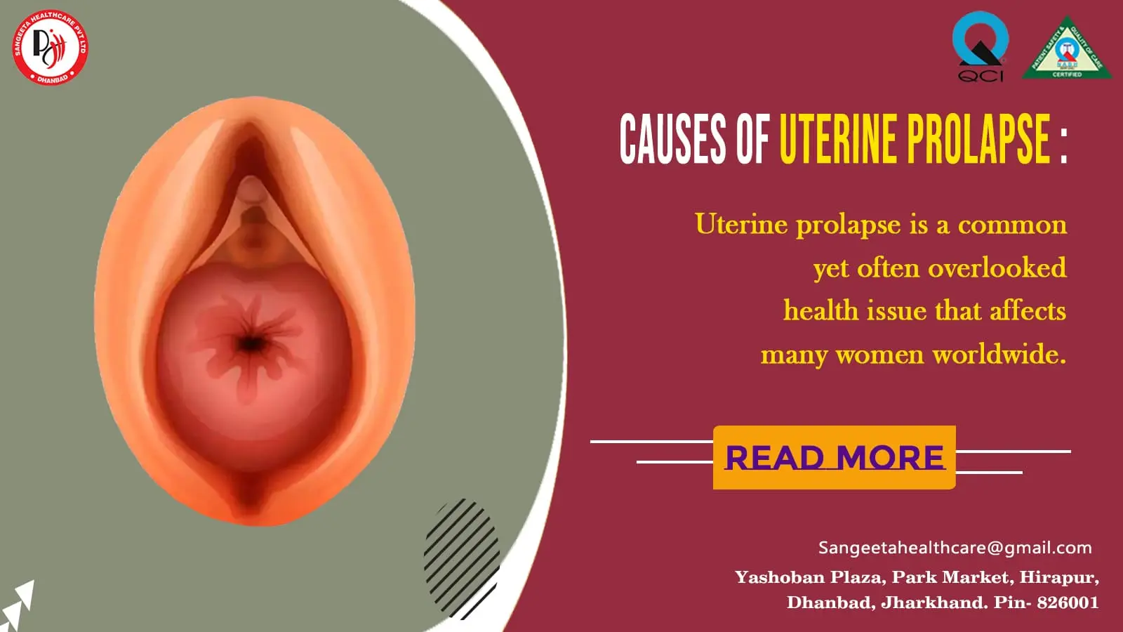
Uterine prolapse is a common yet often overlooked health issue that affects many women worldwide. This condition occurs when the pelvic floor muscles and ligaments weaken, leading to the descent of the uterus into the vaginal canal. In this blog, we will delve into the causes, symptoms, and various treatment options for uterine prolapse, shedding light on a topic that is crucial for women’s health.

Causes of Uterine Prolapse:

  1. Pregnancy and Childbirth: The strain on pelvic muscles during pregnancy and the trauma of childbirth can weaken the supportive tissues of the uterus.
  2. Aging: As women age, hormonal changes and the natural aging process can contribute to the weakening of pelvic floor muscles.
  3. Menopause: Reduced estrogen levels after menopause can lead to a loss of muscle tone and contribute to uterine prolapse.
  4. Hysterectomy: Surgical removal of the uterus can disrupt the supporting structures, increasing the risk of prolapse.
  5. Obesity and Chronic Constipation: Excessive weight and straining during bowel movements can strain the pelvic floor and contribute to prolapse.

Symptoms of Uterine Prolapse:

  1. Sensation of a Bulge: Women with uterine prolapse may feel a bulge or pressure in the vaginal area.
  2. Pelvic Discomfort: Discomfort or pain in the pelvic region, especially when sitting or standing for extended periods.
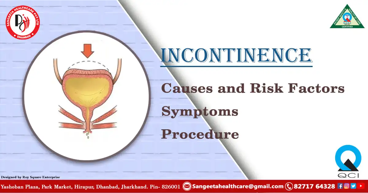
  3. Problems with Urination and Bowel Movements: Difficulty emptying the bladder or bowels, frequent urinary tract infections, and urinary incontinence.
  4. Backache and Lower Abdominal Pain: Pain in the lower back and abdomen may be experienced, particularly during intercourse.
  5. Vaginal Bleeding or Discharge: Some women may notice spotting or an unusual discharge.

Treatment Options:

  1. Pelvic Floor Exercises (Kegels): Strengthening the pelvic floor muscles through exercises can provide support and alleviate mild cases of uterine prolapse.
  2. Hormone Replacement Therapy (HRT): Estrogen therapy may be recommended for postmenopausal women to improve tissue elasticity and muscle tone.
  3. Pessary Use: A pessary is a device inserted into the vagina to support the uterus and alleviate symptoms.
  4. Surgery: In severe cases, surgical interventions like hysterectomy or uterine suspension may be considered to repair and reinforce the pelvic structures.

Prevention and Lifestyle Changes:

1. Maintain a Healthy Weight: Obesity increases the risk of uterine prolapse, so maintaining a healthy weight is crucial.

2. Regular Exercise: Engage in pelvic floor exercises and overall fitness routines to strengthen muscles and maintain a healthy lifestyle.

3. Proper Lifting Techniques: Lift heavy objects using proper techniques to avoid straining the pelvic floor.

Uterine prolapse is a manageable condition with various treatment options available. It is essential for women to be aware of the risk factors, recognize symptoms, and seek timely medical advice. By understanding uterine prolapse, we can empower women to take proactive steps in maintaining their reproductive health and overall well-being.

Uterine prolapse is a condition that can have various implications for a woman’s health, and one of the potential complications associated with it is the development of decubitus ulcers, also known as pressure sores or bedsores. Next, we’ll explore the connection between uterine prolapse and decubitus ulcers, understanding the risk factors, symptoms, prevention, and management strategies.

Understanding Decubitus Ulcers:

Understanding Decubitus Ulcers

Decubitus ulcers are wounds that result from prolonged pressure on the skin, often in areas where bones are close to the skin’s surface. In the context of uterine prolapse, the pressure from the displaced uterus against the vaginal wall and surrounding tissues can create conditions conducive to the development of these ulcers.

Risk Factors:

  1. Prolonged Immobility: Women with uterine prolapse may experience discomfort or pain, leading to reduced mobility, especially if the condition is severe.
  2. Poor Circulation: The pressure exerted by the prolapsed uterus can impede blood flow to specific areas, increasing the risk of tissue damage.
  3. Incontinence: Urinary or fecal incontinence, common in uterine prolapse, can contribute to moisture on the skin, making it more susceptible to breakdown.
  4. Poor Nutrition: Inadequate nutrition can compromise the skin’s integrity, making it more prone to developing ulcers.

Symptoms of Decubitus Ulcers:

  1. Skin Discoloration: Changes in skin color, ranging from red to purple, may be observed.
  2. Swelling and Tenderness: The affected area may become swollen, and the skin may be tender to the touch.
  3. Open Sores or Blisters: As the condition progresses, open sores or blisters may develop, exposing underlying tissues.
  4. Pus or Drainage: Infected ulcers may produce pus or other types of drainage.
  5. Foul Odor: Infections associated with decubitus ulcers can cause a foul odor.

Prevention Strategies:

1. Regular Position Changes: Encourage women with uterine prolapse to change positions frequently to alleviate pressure on vulnerable areas.

2. Use of Supportive Devices: Employing cushions or pillows to support the pelvic region can help distribute pressure more evenly.

3. Maintain Good Hygiene: Regular cleansing and proper care of the perineal area can prevent infections and minimize the risk of ulcers.

4. Nutritional Support: Ensure a well-balanced diet rich in nutrients to promote overall skin health.

5. Seek Prompt Medical Attention: Address any signs of skin breakdown or infection promptly to prevent complications.

Management of Decubitus Ulcers:

  1. Wound Care: Keep the ulcer clean and apply appropriate dressings to promote healing.
  2. Infection Control: Administer antibiotics if there is evidence of infection.
  3. Offloading Pressure: Utilize pressure-relieving devices and techniques to reduce further pressure on affected areas.
  4. Pain Management: Address pain associated with ulcers through medications or alternative pain management strategies.

Understanding the potential risk of decubitus ulcers in the context of uterine prolapse is crucial for both healthcare providers and women themselves. By adopting preventive measures and seeking prompt medical attention, we can minimize the impact of these complications and work towards ensuring the overall well-being of women experiencing uterine prolapse.

Complete procidentia, a severe form of uterine prolapse, poses unique challenges to women’s health, often involving the protrusion of the entire uterus through the vaginal opening. Now, explore the intricacies of complete procidentia and delve into the associated issue of uterine over-distension, shedding light on the causes, symptoms, and potential treatment options.

Understanding Complete Procidentia:

Complete procidentia is an advanced stage of uterine prolapse where the uterus descends entirely into or beyond the vaginal opening. This condition is often characterized by significant discomfort, pain, and a noticeable bulge, impacting a woman’s quality of life and daily activities.

Causes of Complete Procidentia:

  1. Weakened Pelvic Floor Muscles: Prolonged weakening of the pelvic floor muscles, often due to factors like childbirth, aging, or hormonal changes, can contribute to the severity of uterine prolapse.
  2. Multiple Vaginal Deliveries: Women who have undergone multiple vaginal deliveries face an increased risk of complete procidentia.
  3. Hysterectomy Complications: Surgical removal of the uterus (hysterectomy) can sometimes lead to inadequate support of the pelvic structures, increasing the likelihood of prolapse.
  4. Connective Tissue Disorders: Conditions affecting the connective tissues, such as Ehlers-Danlos syndrome, can predispose individuals to uterine prolapse.

Consequences of Uterine Over-Distension:

1. Increased Discomfort and Pain: As the uterus protrudes, it can cause heightened discomfort, pain, and pressure in the pelvic region.

2. Urinary and Fecal Incontinence: Over-distension of the uterus may compromise the integrity of the surrounding structures, leading to issues with bladder and bowel control.

3. Difficulty with Daily Activities: Women with complete procidentia may find it challenging to engage in routine activities such as walking, standing, or even sitting comfortably.

4. Psychological Impact: The physical and emotional toll of complete procidentia can contribute to psychological distress, affecting a woman’s self-esteem and mental well-being.

cause Uterine prolapse

Treatment Options:

  1. Pelvic Floor Exercises (Kegels): Strengthening the pelvic floor muscles can provide support and alleviate mild symptoms.
  2. Pessary Use: A pessary, a device inserted into the vagina to support the uterus, may be recommended to manage symptoms.
  3. Hormone Replacement Therapy (HRT): Estrogen therapy can help improve tissue elasticity and muscle tone, particularly in postmenopausal women.
  4. Surgical Intervention: In severe cases, surgery may be required to repair and reinforce the pelvic structures, possibly involving a hysterectomy.

Lifestyle Changes and Coping Strategies:

  1. Maintain a Healthy Weight: Obesity can exacerbate uterine prolapse, so maintaining a healthy weight is essential.
  2. Avoid Heavy Lifting: Minimize activities that involve heavy lifting to reduce strain on the pelvic floor.
  3. Regular Medical Check-ups: Routine gynecological examinations can help detect and address uterine prolapse at an early stage.

Complete procidentia, coupled with uterine over-distension, demands careful attention and comprehensive management. By understanding the causes, symptoms, and available treatment options, women and healthcare providers can work together to address the challenges posed by this complex condition and improve the overall well-being of those affected. Early intervention and a holistic approach are key to managing complete procidentia and mitigating its impact on women’s lives.


1200x6301.webp
18/Mar/2024

FibroidsFibroids  

Uterine fibroids or Leiomyomas are non-cancerous growths that form inside the uterus. Although often benign, fibroids can cause a range of symptoms and complications for many individuals. In this blog post, we will delve into the causes, symptoms, and treatment options available for fibroids, aiming to provide clarity and understanding for those affected by this condition.

What are Fibroids?

Fibroids are abnormal growths that develop in the muscular wall of the uterus. They vary in size, ranging from tiny seedlings that are undetectable by the human eye to large masses that can distort the shape of the uterus. While the exact cause of fibroids is not fully understood, several factors are believed to contribute to their development, including hormonal imbalances, genetics, and environmental factors.

Symptoms of Fibroids:

The symptoms of fibroids can vary greatly depending on their size, number, and location within the uterus. Some individuals may experience no symptoms at all, while others may encounter significant discomfort and complications.

Common symptoms of fibroids include:

1. Menstrual Irregularities: One of the most common symptoms of fibroids is menstrual irregularities. This can include heavy menstrual bleeding (menorrhagia), prolonged periods, spotting between periods, or irregular menstrual cycles. The presence of fibroids can disrupt the normal functioning of the uterus, leading to changes in the menstrual flow and duration.

2. Pelvic Pain and Pressure: Fibroids can exert pressure on surrounding pelvic organs, leading to pelvic pain and discomfort. Some individuals may experience a dull, aching sensation in the lower abdomen or pelvis, while others may feel sharp or stabbing pains. The severity of pelvic pain can vary depending on the size, number, and location of the fibroids within the uterus.

3. Urinary Symptoms: Fibroids that press against the bladder can cause urinary symptoms such as frequent urination, urgency (sudden and strong urge to urinate), or difficulty emptying the bladder completely. In some cases, fibroids may even lead to urinary retention, where the individual has difficulty urinating altogether.

4. Bowel Symptoms: Fibroids located near the rectum or bowel can exert pressure on these structures, resulting in symptoms such as constipation, difficulty passing stool, or a sensation of rectal fullness. Bowel symptoms associated with fibroids can vary in severity and may worsen during menstruation when fibroids tend to swell due to hormonal changes.

5. Abdominal Enlargement: Large fibroids or multiple fibroids can cause the uterus to enlarge, leading to a noticeable increase in abdominal girth or a feeling of fullness in the abdomen. Abdominal enlargement may be accompanied by other symptoms such as bloating, discomfort, or difficulty fitting into clothing.

6. Pain During Intercourse: Fibroids located near the cervix or within the uterine wall can cause pain or discomfort during sexual intercourse (dyspareunia). This can be due to pressure on sensitive pelvic structures or irritation of the uterine lining caused by friction during intercourse.

Fibroids can manifest in a variety of ways, ranging from subtle discomforts to significant disruptions in daily life. Recognizing the symptoms associated with fibroids is crucial for timely diagnosis and appropriate management. If you’re experiencing any of the symptoms mentioned above or have concerns about your reproductive health, it’s essential to consult with a healthcare provider for further evaluation and guidance. With proper diagnosis and treatment, individuals with fibroids can find relief from their symptoms and improve their overall quality of life.

Diagnosis and Treatment:

Diagnosing fibroids typically involves a combination of medical history review, physical examination, and imaging tests such as ultrasound, MRI, or CT scans. Once diagnosed, treatment options can vary depending on the severity of symptoms, the size and location of the fibroids, and the individual’s reproductive goals.

  1. Watchful Waiting: In cases where fibroids are small and asymptomatic, a “watchful waiting” approach may be recommended. Regular monitoring through exams and imaging can help track any changes in size or symptoms over time.
  2. Medications: Certain medications, such as hormonal birth control or gonadotropin-releasing hormone agonists, may help manage symptoms such as heavy bleeding and pelvic pain by regulating hormonal fluctuations.
  3. Non-Invasive Procedures: Minimally invasive procedures such as uterine artery embolization (UAE) and magnetic resonance-guided focused ultrasound surgery (MRgFUS) can be effective in shrinking fibroids or alleviating symptoms without the need for surgery.
  4. Surgical Intervention: In cases where fibroids are large, causing severe symptoms, or impacting fertility, surgical options such as myomectomy (removal of fibroids while preserving the uterus) or hysterectomy (removal of the uterus) may be considered. 

Different Types of Uterine Fibroid

Leiomyomas, another name for uterine fibroids, are non-cancerous growths that form inside the uterus. While most fibroids are intramural (within the muscular wall of the uterus), there are several distinct types that can develop in different locations. Now, we will explore four specific types of uterine fibroids: broad ligament fibroids, pedunculated fibroids, exophytic fibroids, and parasitic fibroids, shedding light on their characteristics, symptoms, and treatment options.

1. Broad Ligament Fibroids: Broad ligament fibroids are rare and occur within the broad ligament, a band of connective tissue that supports the uterus. These fibroids can grow to a considerable size and may not cause noticeable symptoms until they reach a certain size or position that puts pressure on surrounding organs. Symptoms may include pelvic pain, urinary frequency, and constipation. Treatment options for broad ligament fibroids may include observation, hormonal therapy, or surgical removal, depending on the severity of symptoms and the patient’s reproductive goals.

2. Pedunculated Fibroids: Pedunculated fibroids are fibroids that grow on a stalk (peduncle) attached to the uterine wall. They can either protrude into the uterine cavity (submucosal) or extend outward from the uterine wall (subserosal). Pedunculated fibroids may cause symptoms such as pelvic pain, heavy menstrual bleeding, and pressure on surrounding organs. Treatment options for pedunculated fibroids may include medication to manage symptoms, minimally invasive procedures such as laparoscopic myomectomy to remove the fibroid while preserving the uterus, or hysterectomy in severe cases.

3. Exophytic Fibroids: Exophytic fibroids, also known as subserosal fibroids, grow outward from the outer surface of the uterus. These fibroids may cause symptoms such as pelvic pain, pressure, and a feeling of fullness in the abdomen. While exophytic fibroids may not directly affect fertility, they can impact reproductive health by interfering with the implantation of a fertilized egg or causing complications during pregnancy. Treatment options for exophytic fibroids may include observation, medication to manage symptoms, or surgical removal depending on the severity of symptoms and the patient’s reproductive goals.

4. Parasitic Fibroids: Parasitic fibroids are a rare type of fibroid that occurs when a fibroid loses its blood supply from the uterus and attaches to another organ, such as the ovaries or the abdominal wall, for a continued blood supply. These fibroids may cause symptoms such as pelvic pain, abdominal swelling, and urinary or bowel symptoms. Treatment for parasitic fibroids typically involves surgical removal to alleviate symptoms and prevent complications.

Uterine fibroids come in various forms, each with its own characteristics, symptoms, and treatment considerations. Broad ligament fibroids, pedunculated fibroids, exophytic fibroids, and parasitic fibroids present unique challenges in diagnosis and management. Individuals experiencing symptoms suggestive of uterine fibroids should seek medical evaluation for proper diagnosis and personalized treatment planning. With advancements in medical technology and a comprehensive understanding of fibroid types, individuals can receive appropriate care to address their symptoms and improve their quality of life.

Fibroids size

Understanding Fibroid Sizes: When Should You Be Concerned?


Fibroids, non-cancerous growths that develop in the uterus, come in various sizes and can affect individuals differently. While many fibroids cause no symptoms and are harmless, others can lead to discomfort, complications, and health risks. In this blog post, we’ll explore the different sizes of fibroids and when they might pose a concern for your health.

Small Fibroids:

Small fibroids, typically less than 3 centimeters (about 1.2 inches) in diameter, are often asymptomatic and may not require treatment. These tiny fibroids are usually detected incidentally during routine pelvic exams or imaging studies for other reasons. While small fibroids may cause minimal discomfort, they generally don’t pose significant health risks.

Medium-Sized Fibroids:

Medium-sized fibroids range from 3 to 10 centimeters (about 1.2 to 3.9 inches) in diameter. While they may still be asymptomatic in some cases, medium-sized fibroids can cause symptoms such as heavy menstrual bleeding, pelvic pain, pressure on the bladder or bowel, and fertility issues. Depending on their location and symptoms, treatment options for medium-sized fibroids may include medication, minimally invasive procedures, or surgery.

Large Fibroids:

Large fibroids, measuring over 10 centimeters (about 3.9 inches) in diameter, are more likely to cause noticeable symptoms and complications. These fibroids can distort the shape of the uterus, cause significant pelvic pain and pressure, and lead to complications such as anemia due to heavy menstrual bleeding. Large fibroids may also interfere with fertility and increase the risk of pregnancy complications. Treatment for large fibroids may involve medication to manage symptoms, minimally invasive procedures to shrink the fibroids, or surgical removal, depending on the severity of symptoms and the patient’s reproductive goals.

When Are Fibroids Dangerous?

While fibroids of any size can cause discomfort and complications, certain factors may indicate a higher risk of complications:

1.Rapid Growth: Fibroids that grow rapidly may indicate a more aggressive type or an underlying hormonal imbalance, requiring closer monitoring and evaluation.
2.Location: Fibroids located near critical structures such as the bladder or bowel may cause more pronounced symptoms and complications.
3.Pregnancy: Fibroids that increase in size during pregnancy or cause complications such as miscarriage, preterm labor, or abnormal fetal position may require prompt medical attention.
4.Severe Symptoms: Fibroids causing severe symptoms such as intense pelvic pain, significant blood loss, or urinary/bowel dysfunction may necessitate immediate medical intervention.

Fibroids Dangerous

While fibroids of any size can impact an individual’s health and well-being, larger fibroids and those causing severe symptoms or complications may require medical evaluation and treatment. If you’re experiencing symptoms suggestive of fibroids or have concerns about your reproductive health, it’s essential to consult with a healthcare provider for proper diagnosis and personalized treatment recommendations. With timely intervention and appropriate management, individuals with fibroids can find relief from their symptoms and improve their quality of life.

Fibroids are a common gynecological condition that can significantly impact a person’s quality of life. While they are usually benign, the symptoms they cause can be disruptive and distressing. It’s essential for individuals experiencing symptoms of fibroids to seek medical evaluation and explore treatment options tailored to their unique circumstances. With proper diagnosis and management, individuals with fibroids can find relief from their symptoms and regain control of their health and well-being.


1200x630-2-1.webp
11/Mar/2024

Peptic Ulcer Disease

Peptic Ulcer Disease

Ulcers are common afflictions that can affect various parts of the digestive system, each with its distinct characteristics and causes. Peptic Ulcer Disease (PUD) is a prevalent gastrointestinal disorder that affects millions of people worldwide. In this blog post, we will delve into the intricacies of peptic ulcers, exploring their causes, symptoms, and available treatments to provide a comprehensive understanding of this condition.

What is Peptic Ulcer Disease?

Peptic Ulcer Disease refers to the formation of open sores or ulcers on the inner lining of the stomach, upper small intestine, or esophagus. These ulcers develop when the protective lining of the digestive tract is compromised, allowing stomach acid to erode the underlying tissues.

Causes:

• Helicobacter pylori Infection : The majority of peptic ulcers are associated with an infection caused by the bacterium Helicobacter pylori (H. pylori). This bacterium weakens the stomach’s protective mucus layer, making it susceptible to damage from stomach acid.
• Nonsteroidal Anti-Inflammatory Drugs (NSAIDs) : Regular use of NSAIDs, such as aspirin and ibuprofen, can contribute to the development of peptic ulcers. These medications can irritate the stomach lining and increase the risk of ulcer formation.
• Acid Production : Excessive stomach acid production can also be a contributing factor. This may occur due to various reasons, including stress, smoking, and certain dietary habits.

Symptoms:

The symptoms of peptic ulcers can vary, and some individuals may experience no symptoms at all. Common signs include:
Burning stomach pain: This pain is often felt between meals or during the night.
Nausea and vomiting: Some individuals may experience nausea and vomiting, especially after meals.
Bloating and belching: Excessive gas and belching can be indicative of peptic ulcers.
Unexplained weight loss: In severe cases, peptic ulcers may lead to unintended weight loss.

Pathogenesis of Peptic Ulcer Disease

The pathogenesis of peptic ulcer disease (PUD) involves a complex interplay of factors that ultimately lead to the formation of open sores or ulcers in the gastrointestinal tract. The primary site for peptic ulcers is the stomach and the upper part of the small intestine (duodenum). The key factors contributing to the pathogenesis of peptic ulcer include:

1. Helicobacter pylori Infection:
  • The bacterium Helicobacter pylori (H. pylori) is a major contributor to peptic ulcer development.
  • H. pylori infection weakens the protective mucous layer of the stomach, making the underlying tissues more susceptible to damage from stomach acid.
  • The bacterium can directly damage the lining of the stomach and duodenum, leading to inflammation and ulceration.
2. Increased Acid Production:

• Excessive production of stomach acid, known as hyperacidity, can contribute to the erosion of the protective mucosal barrier.
• Factors such as stress, certain dietary habits, and smoking can stimulate the production of gastric acid, creating an environment conducive to ulcer formation.

3. Use of Nonsteroidal Anti-Inflammatory Drugs (NSAIDs):

• Regular use of NSAIDs, including aspirin, ibuprofen, and naproxen, is a well-established risk factor for peptic ulcers.
• NSAIDs can irritate the stomach lining and impair the protective mechanisms, leading to increased susceptibility to ulcer formation.

4. Impaired Mucosal Defense Mechanisms:

• The stomach and duodenum have protective mechanisms to prevent damage from the acidic environment. Disruption of these mechanisms can contribute to ulcer development.
• Factors such as a decrease in mucous production, impaired blood flow to the mucosa, or a reduction in the ability to repair damaged tissue can compromise the mucosal defense.

5. Genetic Factors:

• There is evidence to suggest that genetic factors may play a role in predisposing individuals to peptic ulcer disease.
• Some people may have a genetic predisposition to an exaggerated response to H. pylori infection or an increased susceptibility to environmental factors that contribute to ulceration.

6. Stress and Lifestyle Factors:

• While stress alone is not a direct cause of peptic ulcers, it can exacerbate existing conditions and contribute to the development of ulcers in susceptible individuals.
• Lifestyle factors such as poor dietary choices, smoking, and excessive alcohol consumption can also contribute to the pathogenesis of peptic ulcers.

Diagnosis:

Diagnosing peptic ulcer disease typically involves a combination of medical history analysis, physical examination, and diagnostic tests. These tests may include endoscopy, blood tests, and imaging studies to determine the presence and severity of ulcers.

Treatment:
  1. Antibiotics: If H. pylori infection is identified, a course of antibiotics is prescribed to eradicate the bacteria.
  2. Acid-suppressing medications: Proton pump inhibitors (PPIs) and histamine-2 (H2) blockers help reduce stomach acid production, allowing the ulcers to heal.
  3. Antacids: These provide quick relief by neutralizing stomach acid.
  4. Lifestyle changes: Avoiding NSAIDs, managing stress, quitting smoking, and making dietary modifications can contribute to the healing process.

Understanding Benign Gastric Ulcers

gastric ulcers

Gastric ulcers are localized disruptions in the stomach lining, and when they are benign, it means they are not cancerous. These ulcers can range in size and severity, causing discomfort and potential complications if left untreated.

Causes:

1.Helicobacter pylori Infection: One of the primary causes of benign gastric ulcers is infection with the bacterium Helicobacter pylori (H. pylori). This bacterium weakens the protective mucous layer in the stomach, making the lining susceptible to damage from stomach acid.
2.Nonsteroidal Anti-Inflammatory Drugs (NSAIDs): Regular use of NSAIDs, such as aspirin or ibuprofen, is a common risk factor for benign gastric ulcers. These medications can irritate the stomach lining, leading to ulcer formation.
3.Excessive Stomach Acid: An overproduction of stomach acid can contribute to the development of gastric ulcers. This may result from stress, certain dietary habits, or other factors that stimulate acid secretion.
4.Smoking: Smoking has been identified as a risk factor for gastric ulcers. It may impair the healing of existing ulcers and increase susceptibility to developing new ones.

Symptoms:

The symptoms of benign gastric ulcers can vary, and some individuals may experience no symptoms at all. Common signs include:
•Burning or gnawing stomach pain: This pain is often felt in the upper abdomen, typically between meals or during the night.
•Nausea and vomiting: Some individuals may experience nausea and vomiting, especially after meals.
•Bloating and belching: Excessive gas and belching can occur.
•Unexplained weight loss: In more severe cases, gastric ulcers may lead to unintended weight loss.

Diagnosis: Diagnosing benign gastric ulcers involves a combination of medical history analysis, physical examination, and diagnostic tests. Tests may include endoscopy, blood tests, and imaging studies to confirm the presence and severity of ulcers.

Understanding Liver Ulcers:

The liver, a vital organ with multifaceted functions, is susceptible to various disorders. One such condition that warrants attention is liver ulcers. Now, we will discuss intricacies of liver ulcers, exploring their causes, symptoms, and potential approaches to care. Liver ulcers, also known as hepatic ulcers, refer to open sores or lesions on the liver’s surface. While less common than other liver disorders, such as cirrhosis or hepatitis, liver ulcers can pose serious health concerns and require careful management.

Causes:

•Infections: Certain infections, including parasitic, bacterial, or fungal infections, can lead to the development of liver ulcers. These infections may compromise the liver’s protective mechanisms, allowing ulcers to form.
•Trauma: Physical trauma or injury to the liver, whether due to accidents, surgery, or other external factors, can contribute to ulcer formation.
•Vascular Issues: Conditions affecting blood vessels around the liver, such as Budd-Chiari syndrome, can lead to reduced blood flow, causing ulcers.
•Autoimmune Diseases: Disorders where the immune system mistakenly attacks liver tissues can increase the risk of ulcer development.

Symptoms:

Liver ulcers may present with a range of symptoms, and their severity can vary. Common signs include:
•Abdominal pain: Persistent or sharp pain in the upper right abdomen.
•Jaundice: Yellowing of the skin and eyes due to impaired liver function.
•Fatigue: Generalized weakness and tiredness.
•Unexplained weight loss: Loss of weight without an apparent cause.

liver ulcer

Diagnosis:

Diagnosing liver ulcers involves a combination of medical history assessment, physical examination, and diagnostic tests. Imaging studies such as ultrasound, CT scans, or MRI may be employed to visualize the liver and identify ulcers.

Treatment:

•Antibiotics: If the ulcers are caused by bacterial infections, a course of antibiotics is often prescribed.
•Antifungal or Antiparasitic Medications: In the case of fungal or parasitic infections, specific medications may be recommended.
•Pain Management: Analgesics or anti-inflammatory medications may be prescribed to alleviate pain.
•Immune System Modulators: For autoimmune-related liver ulcers, medications that modulate the immune response may be prescribed.
•Surgery: In severe cases or when complications arise, surgical intervention may be necessary to address the ulcers.

Burn Ulcers & Antral Ulcers

Apart from liver ulcers and peptic ulcers , burn ulcers and antral ulcers are two other common types of ulcers. Next, we will delve into two specific types of ulcers – burn ulcers and antral ulcers – exploring their unique features, causes, and potential treatments.

Understanding Burn Ulcers:

Burn ulcers, often referred to as stress ulcers, are a type of gastrointestinal ulcer that can occur following severe physical injury or trauma, such as extensive burns, major surgery, or severe infections. These ulcers are believed to be triggered by a combination of factors, including reduced blood flow to the stomach lining, a compromised mucous barrier, and increased stomach acid production due to stress.

Causes of Burn Ulcers:

•Severe Physical Trauma: Burn ulcers are commonly associated with significant physical trauma, such as burns covering a large surface area.
•Surgery: Major surgical procedures, especially those involving the abdomen, can contribute to the development of burn ulcers.
•Sepsis or Infections: Severe infections and conditions like sepsis can increase the risk of burn ulcers.

Symptoms of Burn Ulcers:

•Abdominal Pain: Upper abdominal ache that never goes away.
•Nausea and Vomiting: Individuals may experience nausea and vomiting, especially after meals.
•Bleeding: In severe cases, burn ulcers can lead to gastrointestinal bleeding.

Treatment of Burn Ulcers:

•Stress Management: Reducing stress and anxiety through relaxation techniques can be beneficial.
•Acid-Suppressing Medications: Proton pump inhibitors (PPIs) or histamine-2 (H2) blockers may be prescribed to decrease stomach acid production.
•Mucosal Protectants: Medications that enhance the protective mucous layer of the stomach may be used.

Understanding Antral Ulcers:

Antral ulcers specifically refer to ulcers that develop in the antrum, which is the lower portion of the stomach. These ulcers are often associated with Helicobacter pylori (H. pylori) infection and disturbances in the regulation of stomach acid.

Causes of Antral Ulcers:

•H. pylori Infection: The bacterium H. pylori is a major contributor to antral ulcers by compromising the protective mucous layer.
•Increased Stomach Acid: Factors leading to elevated stomach acid production can contribute to antral ulcer development.
•NSAID Use: Regular use of nonsteroidal anti-inflammatory drugs (NSAIDs) can irritate the stomach lining and increase the risk of antral ulcers.

Symptoms of Antral Ulcers:

•Burning or Gnawing Pain: Pain in the upper abdomen, often between meals or during the night.
•Nausea and Bloating: Antral ulcers may cause symptoms of nausea and bloating.
•Unexplained Weight Loss: In severe cases, weight loss may occur.

Treatment of Antral Ulcers:

•Antibiotics: If H. pylori infection is confirmed, a course of antibiotics is prescribed to eliminate the bacteria.
•Acid-Suppressing Medications: PPIs and H2 blockers help reduce stomach acid production.
•Antacids: They neutralize stomach acid and offer immediate relief.

While different types of ulcers share some common symptoms, their causes and treatment approaches differ. Understanding the distinctive features of each type of ulcer is crucial for accurate diagnosis and effective management. If you suspect you have ulcers or are experiencing persistent symptoms, seek professional medical advice for a thorough evaluation and personalized treatment plan tailored to your specific needs. Early intervention can lead to better outcomes and improved gastrointestinal health.


1200x630-2.webp
04/Mar/2024

Millions of women worldwide suffer with the persistent and frequently incapacitating disease known as endometriosis.Despite its prevalence, the awareness surrounding endometriosis remains limited, leading to delayed diagnoses and a lack of understanding about the impact it can have on a person’s life. In this blog post, we’ll delve into the intricacies of endometriosis, exploring its definition, symptoms, diagnosis, and the challenges faced by those living with this condition.

endometriosis

What is Endometriosis?

Endometriosis is a medical condition where tissue similar to the lining of the uterus, known as the endometrium, grows outside the uterus. This misplaced tissue can be found on the ovaries, fallopian tubes, bladder, and other pelvic organs. Unlike the normal endometrial tissue, the displaced tissue has no way to exit the body, leading to inflammation, pain, and the formation of scar tissue.

Symptoms:

The symptoms of endometriosis can vary widely, making it challenging to diagnose. Common symptoms include:

1. Pelvic Pain: Persistent and often severe pelvic pain is a hallmark of endometriosis. The pain may occur before or during menstruation, during sexual intercourse, or even during bowel movements.

2. Menstrual Irregularities: Women with endometriosis may experience heavy or irregular periods.

3. Painful Intercourse: Endometriosis can cause deep pain during sexual intercourse, which can significantly impact a person’s quality of life and relationships.

4. Digestive Issues: Some individuals may experience gastrointestinal symptoms such as bloating, constipation, or diarrhea, especially during menstruation.

Uncommon symptoms of endometriosis

However, what often goes unnoticed are the less common symptoms that can significantly impact a person’s life. Here, we will explore some of these uncommon symptoms, shedding light on the diverse ways endometriosis can manifest.

1. Chronic Fatigue:

While fatigue is a common complaint in many medical conditions, those with endometriosis often experience an overwhelming and persistent fatigue that goes beyond the usual tiredness associated with a hectic lifestyle. The chronic inflammation and hormonal imbalances caused by endometriosis may contribute to this profound fatigue, affecting daily activities and overall quality of life.

2. Painful Bowel Movements:

Endometriosis can involve the bowels, leading to pain and discomfort during bowel movements. Some individuals may experience diarrhea, constipation, or a mix of both, particularly around the time of menstruation. This symptom is often overlooked or attributed to other gastrointestinal issues, delaying the diagnosis of endometriosis.

3. Painful Urination:

Endometrial tissue may implant itself on the bladder or urinary tract, causing pain and discomfort during urination. This symptom can be mistaken for a urinary tract infection, leading to repeated courses of antibiotics without relief.

4. Painful Ovulation:

Women with endometriosis may experience sharp pain or cramping during ovulation, making it uncomfortable and sometimes debilitating. This symptom can be mistaken for normal menstrual cramps, resulting in a lack of recognition and appropriate management.

5. Pain Beyond the Pelvis:

While pelvic pain is a hallmark of endometriosis, the pain can extend beyond the pelvic region. Some individuals may experience pain in the lower back, thighs, or even radiating down the legs. This widespread pain can be challenging to connect with endometriosis, leading to a delay in diagnosis.

6. Cyclical Migraines:

Hormonal fluctuations associated with the menstrual cycle can trigger migraines in individuals with endometriosis. These cyclical migraines may be intense and challenging to manage, impacting daily functioning during menstruation.Understanding that endometriosis is not solely defined by pelvic pain and menstrual irregularities is crucial for timely diagnosis and effective management. By recognising the less common symptoms, individuals, healthcare providers, and the general public can contribute to a broader understanding of endometriosis. If you or someone you know is experiencing any of these unusual symptoms, seeking medical attention and advocating for comprehensive evaluation can make a significant difference in managing the impact of endometriosis on daily life. Let’s bring these lesser-known aspects of endometriosis into the spotlight and work towards a more informed and supportive approach to this complex condition.

Diagnosis:

Diagnosing endometriosis can be challenging, and it often takes several years from the onset of symptoms for a correct diagnosis to be made. Common diagnostic methods include:

1.Medical History and Symptom Assessment: A thorough discussion of symptoms, their duration, and their impact on daily life is crucial.
2. Physical Examination: A pelvic examination may be performed to check for abnormalities or signs of endometriosis.
3. Imaging Studies: Ultrasound or MRI scans may be conducted to identify the presence of endometrial tissue outside the uterus.
4. Laparoscopy:The most definitive method of diagnosis involves a surgical procedure called laparoscopy, where a small camera is inserted into the pelvic area to visualize and confirm the presence of endometrial tissue.

Challenges and Impact:

Endometriosis can have a profound impact on a person’s physical, emotional, and social well-being. Some of the challenges faced by those with endometriosis include:

1. Misunderstanding and Stigma: Due to a lack of awareness, there is often a misunderstanding of endometriosis, leading to stigmatization of those affected.
2. Delayed Diagnosis: The vague and diverse nature of symptoms contributes to delays in diagnosis, impacting treatment outcomes.
3. Fertility Issues: Endometriosis is a common cause of infertility, adding an additional layer of complexity and emotional distress for those trying to conceive.

Understanding Bowel Endometriosis:

Bowel endometriosis occurs when endometrial-like tissue implants itself on the bowel or adjacent structures. This can lead to various symptoms, including abdominal pain, bloating, changes in bowel habits, and discomfort during bowel movements. While these symptoms alone can significantly affect a person’s quality of life, the implications of bowel endometriosis extend to fertility concerns as well.

Impact on Fertility:

Adhesions and Scar Tissue:

Bowel endometriosis can cause the formation of adhesions and scar tissue in the pelvic region. These adhesions may distort the anatomy of reproductive organs, affecting the normal function of the fallopian tubes and ovaries. The resulting mechanical obstacles can impede the journey of the egg and sperm, reducing the chances of conception.

Inflammation and Hormonal Imbalance:

The inflammatory response triggered by endometriosis, particularly in the bowel, can create a hostile environment for reproductive organs. This chronic inflammation and hormonal imbalances may disrupt the delicate hormonal interplay necessary for a healthy menstrual cycle and successful conception.

Surgical Interventions:

In cases of severe bowel endometriosis, surgical interventions may be necessary to remove the affected tissue and alleviate symptoms. However, these surgeries can also pose a risk to fertility, as they may involve the removal of reproductive organs or the formation of additional scar tissue.

Navigating Fertility Challenges:

Early Diagnosis and Intervention:

Early diagnosis of bowel endometriosis is crucial for managing both symptoms and fertility concerns. Seeking medical attention when experiencing symptoms and undergoing comprehensive evaluations, including imaging studies and laparoscopy, can aid in timely intervention.

Collaborative Approach:

Managing bowel endometriosis and its impact on fertility often requires a collaborative approach involving gynecologists, gastroenterologists, and fertility specialists. A multidisciplinary team can tailor a treatment plan that addresses both the gastrointestinal and reproductive aspects of the condition.

Fertility Preservation Options:

For individuals facing fertility challenges due to bowel endometriosis, fertility preservation options such as egg freezing or in vitro fertilization (IVF) may be considered. These approaches aim to secure the possibility of conception before undergoing treatments that might impact fertility.Increased awareness, early diagnosis, and effective management are crucial for addressing the challenges posed by endometriosis. By fostering understanding and support, we can empower individuals with endometriosis to navigate their journey towards improved health and quality of life. It’s time to shed light on this often overlooked condition and work towards a future where those affected by endometriosis receive the care, understanding, and support they deserve.


Pelvic-Inflammatory-Disease-blog-post12006301.webp
26/Feb/2024

Pelvic Inflammatory Disease

Pelvic Inflammatory Disease (PID)

Pelvic Inflammatory Disease (PID) is a serious health condition that affects the female reproductive system. It occurs when bacteria from the vagina or cervix travel upwards into the uterus, fallopian tubes, or ovaries, causing inflammation and infection. PID can have severe consequences if left untreated, including infertility, chronic pelvic pain, and an increased risk of ectopic pregnancy. In this blog post, we will explore the causes, symptoms, and treatment options for Pelvic Inflammatory Disease.

Causes:

The primary cause of PID is the introduction of bacteria into the upper genital tract. The most common bacteria responsible for PID are sexually transmitted infections (STIs), such as Chlamydia and Gonorrhea. However, other bacteria not related to sexual activity can also contribute to PID. Risk factors for PID include multiple sexual partners, a history of STIs, and the use of intrauterine devices (IUDs) for contraception.

Symptoms:

PID symptoms can vary in severity, and some individuals may experience mild discomfort while others may have more pronounced symptoms. Common signs of PID include:

  • Pelvic Pain: Persistent and dull pain in the lower abdomen and pelvic region.
  • Abnormal Vaginal Discharge: Unusual discharge that may be yellow or green with a foul odor.
  • Painful Urination: Discomfort or pain while urinating.
  • Irregular Menstrual Bleeding: Changes in the menstrual cycle, including heavier or more painful periods.
  • Fever and Chills: Elevated body temperature and chills, indicating an infection.

It’s important to note that some women with PID may not exhibit any noticeable symptoms, making regular gynecological check-ups crucial for early detection.

Complications:

If left untreated, Pelvic Inflammatory Disease can lead to serious complications, including:

  1. Infertility: Scar tissue from the inflammation can block the fallopian tubes, preventing the egg from reaching the uterus.
  2. Chronic Pelvic Pain: Persistent pain in the pelvic region that may last for months or years.
  3. Ectopic Pregnancy: The risk of an ectopic pregnancy (a pregnancy outside the uterus) is higher for women who have had PID.

Treatment:

Prompt and appropriate treatment is crucial to prevent complications. Treatment typically involves a combination of antibiotics to eliminate the infection. In severe cases or if an abscess has formed, hospitalization may be necessary for intravenous antibiotics and, in some cases, surgical intervention to drain the abscess.

Prevention:

Preventing PID involves practicing safe sex, using barrier methods of contraception, and getting regular screenings for STIs. If diagnosed with an STI, it is essential to complete the prescribed course of antibiotics and inform sexual partners to prevent the spread of infection.

Reproductive Tract Infection (RTI)

Reproductive Tract Infection

When it comes to the health of kidney, RTI is the most common terms that affects the organ irrespective of age and gender.

Understand the term:

A Reproductive Tract Infection (RTI) refers to an infection that affects the reproductive organs and structures in both men and women. These infections can involve various components of the reproductive system, including the uterus, fallopian tubes, ovaries, cervix, vagina in women, and the testes, prostate, and other related structures in men. RTIs can be caused by a variety of microorganisms, including bacteria, viruses, fungi, and parasites.

Classification:

RTIs can be classified into two main categories:

Sexually Transmitted Infections (STIs): These are infections that are primarily transmitted through sexual contact. Common STIs that can affect the reproductive tract include Chlamydia, Gonorrhea, Syphilis, Herpes, and Human Papillomavirus (HPV).

Non-Sexually Transmitted Infections: These infections may occur due to non-sexual factors such as poor hygiene, compromised immune function, or the presence of other medical conditions. Bacterial vaginosis, yeast infections, and urinary tract infections are examples of non-sexually transmitted reproductive tract infections.

Symptoms& Prevention:

The symptoms of RTIs can vary widely depending on the specific infection, its severity, and the affected individual’s gender. Common symptoms may include abnormal vaginal or penile discharge, pain or discomfort during urination, pelvic pain, lower abdominal pain, and changes in menstrual patterns.

It’s crucial to diagnose and treat RTIs promptly to prevent complications such as pelvic inflammatory disease, infertility, ectopic pregnancies, and an increased risk of HIV transmission. Prevention measures include practicing safe sex, maintaining good personal hygiene, regular medical check-ups, and, in the case of STIs, informing and treating sexual partners.

As with any medical concern, individuals experiencing symptoms associated with reproductive tract infections should seek medical attention for an accurate diagnosis and appropriate treatment. Early intervention can help manage these infections effectively and reduce the risk of long-term complications.

Minimal pod fluid

In the realm of medical diagnostics, minimal pod fluid is another term that may surface during various tests, particularly when assessing the health of the kidneys. Understanding what minimal pod fluid entails is crucial for individuals and healthcare providers alike. Nextin  this blog post, we’ll explore the concept of minimal pod fluid, its significance, and what it might indicate for your health.

Defining Minimal Pod Fluid:

Minimal pod fluid, also known as minimal change disease (MCD), refers to a kidney condition characterized by abnormal functioning of the glomeruli, the tiny filtering units within the kidneys. In individuals with minimal pod fluid, these glomeruli appear normal under a microscope, and the condition is often diagnosed through a renal biopsy.

Causes:

The exact cause of minimal pod fluid is not always clear, but it is believed to be related to an immune system dysfunction. In some cases, it may be triggered by infections, allergic reactions, or certain medications. Minimal change disease is more commonly observed in children, but it can affect individuals of any age.

Symptoms:

One of the challenging aspects of minimal pod fluid is that it often presents with few or no noticeable symptoms. However, some individuals may experience:

  • Swelling (Edema): Swelling, particularly in the legs and around the eyes, may occur due to the retention of fluids.
  • Foamy Urine: The presence of excess protein in the urine can result in foamy or bubbly urine.
  • Fatigue:It’s possible for some people to feel weak or exhausted.

Diagnosis:

The diagnosis of minimal pod fluid typically involves a combination of medical history, physical examination, and laboratory tests. A renal biopsy is often the definitive method to confirm the presence of minimal change disease and rule out other kidney conditions.

Treatment:

The good news is that minimal pod fluid often responds well to treatment. The primary approach involves the use of corticosteroid medications, which can help reduce inflammation and improve kidney function. In most cases, individuals with minimal change disease have a favourable prognosis, with many achieving complete remission.

While minimal pod fluid might sound concerning, it is important to remember that it is a treatable condition, especially when diagnosed early. Regular check-ups, particularly if you notice any unusual symptoms like swelling or changes in urine, are essential for maintaining kidney health. If you or someone you know is diagnosed with minimal pod fluid, working closely with healthcare providers to develop a personalized treatment plan can lead to successful management and improved long-term outcomes.

Inflammatory Smear:

The realm of women’s health is vast and intricate, with various screenings and tests aimed at maintaining reproductive well-being. One such crucial diagnostic tool is the inflammatory smear, an examination that plays a pivotal role in assessing the health of the female reproductive system. Now, we’ll delve into the significance of inflammatory smears, what they entail, and why they are an essential aspect of women’s healthcare.

Understanding Inflammatory Smear:

An inflammatory smear, commonly known as a Pap smear or Pap test, is a routine screening procedure designed to detect abnormal changes in the cells of the cervix, the lower part of the uterus. The primary goal of this test is to identify early signs of cervical cancer or precancerous conditions, allowing for timely intervention and prevention.

Procedure:

During the inflammatory smear procedure, a healthcare provider collects a small sample of cells from the cervix using a soft brush or spatula. The collected cells are then examined under a microscope to identify any abnormalities, such as changes in size, shape, or organization. This microscopic analysis helps detect potential issues at an early stage when they are more manageable and treatable.


Importance of Inflammatory Smear:

Understanding Inflammatory Smear

  • Early Detection of Cervical Abnormalities: Inflammatory smears are instrumental in identifying abnormal cellular changes in the cervix, which could indicate the presence of human papillomavirus (HPV) or other factors that may lead to cervical cancer.
  • Preventing Cervical Cancer: By detecting and addressing abnormalities early, inflammatory smears contribute significantly to preventing the development of cervical cancer. Early intervention can involve monitoring, medical treatments, or minor surgical procedures, depending on the severity of the findings.
  • Monitoring Reproductive Health: Inflammatory smears are part of routine gynaecological examinations, allowing healthcare providers to monitor a woman’s reproductive health over time. Regular screenings help track changes, assess risk factors, and tailor preventive measures accordingly.
  • HPV Detection: Inflammatory smears also play a crucial role in detecting the presence of HPV, a common sexually transmitted infection linked to cervical cancer. Identifying HPV early enables healthcare providers to monitor and manage the infection effectively.

Inflammatory smears are an indispensable tool in women’s healthcare, contributing to the prevention and early detection of cervical abnormalities. Regular screenings empower women to take charge of their reproductive health, providing a foundation for timely interventions that can ultimately save lives. It’s essential for women to prioritize routine gynaecological check-ups, including inflammatory smears, as part of a proactive approach to overall well-being. Remember, early detection is key, and staying informed about your health is a powerful step towards a healthier future.

In the conclusion of Pelvic Inflammatory Disease, it can be said that it is a serious health concern that requires prompt attention. Understanding the causes, recognizing symptoms, and seeking timely medical intervention are crucial for preventing long-term complications. Regular gynaecological check-ups, open communication with healthcare providers, and practicing safe sex are essential steps in maintaining reproductive health and preventing the occurrence of PID.


1200x630-1-1-3.webp
19/Feb/2024

Incontinence


Incontinence
Living with incontinence can be a challenging and sensitive experience for individuals of all ages. It’s a condition that often goes undiscussed due to the stigma attached to it. In this blog post, we aim to shed light on incontinence, its causes, types, and management strategies, while promoting understanding, empathy, and support for those dealing with this common yet often misunderstood condition.

Understanding Incontinence:

Incontinence refers to the inability to control the release of urine or faeces, leading to involuntary leakage. This condition can affect people of all ages, genders, and backgrounds, and it may be a symptom of an underlying medical issue.

Types of Incontinence:

• Stress Incontinence : Caused by pressure on the bladder, such as coughing, sneezing, or physical activity.
• Urge Incontinence : Involves a sudden and intense urge to urinate, often resulting in leakage.
• Overflow Incontinence : Occurs when the bladder overflows due to improper emptying.
• Functional Incontinence : Not related to bladder or urinary dysfunction but rather to physical or cognitive limitations that hinder reaching the bathroom in time.

Causes and Risk Factors:

Understanding the root causes of incontinence is crucial for effective management. Common factors include age, pregnancy and childbirth, obesity, neurological conditions, and certain medications. It’s essential to identify and address these underlying issues to tailor an appropriate treatment plan.

Management Strategies:

• Lifestyle Changes : Adopting a healthy lifestyle, including a balanced diet, regular exercise, and maintaining a healthy weight, can significantly improve incontinence symptoms.
• Pelvic Floor Exercises : Kegel exercises help strengthen the pelvic floor muscles, providing better control over bladder function.
• Medications : Depending on the type of incontinence, medications may be prescribed to manage symptoms.
• Behavioural Therapies : Techniques such as bladder training, scheduled voiding, and fluid management can be effective in improving control.
• Medical Devices : In some cases, medical devices such as catheters or pessaries may be recommended for managing incontinence.

Psychosocial Impact:

Beyond the physical aspects, incontinence can have a profound impact on an individual’s mental and emotional well-being. It’s crucial to address the psychosocial aspects of incontinence, offering support, understanding, and resources for coping with the challenges it presents.

Reflex incontinence

1. reflex incontinence

Reflex incontinence is a specific type of urinary incontinence that often goes undiscussed, yet it can significantly affect an individual’s daily life. Next in this blog post, we will delve into the world of reflex incontinence, exploring its causes, symptoms, and potential management strategies to empower those grappling with this condition.

Defining Reflex Incontinence:

Reflex incontinence, also known as neurogenic bladder, is characterized by the inability to control the release of urine due to a lack of awareness or control over bladder function. Unlike other types of incontinence, reflex incontinence is associated with neurological issues that disrupt the normal signals between the bladder and the brain.

Causes of Reflex Incontinence:

•  Spinal Cord Injury : Trauma to the spinal cord can interfere with nerve signals responsible for controlling bladder function.
•  Neurological Diseases : Conditions such as multiple sclerosis, Parkinson’s disease, or stroke can impact the nerves controlling the bladder.
•  Spinal Cord Tumors : Growths or tumors on the spinal cord may disrupt normal nerve communication, leading to reflex incontinence.
•  Diabetic Neuropathy : Diabetes can cause nerve damage, including those responsible for bladder control.

Symptoms:

The primary symptom of reflex incontinence is the involuntary release of urine without the individual being consciously aware or in control. This often happens suddenly and may be triggered by a reflex, such as when the bladder is full.

Challenges and Impact:

Living with reflex incontinence presents unique challenges. Individuals may face difficulties in social situations, experience embarrassment, and struggle with a reduced quality of life. Addressing these challenges requires a holistic approach that encompasses both physical and emotional well-being.

Management Strategies:

•  Catheterization : Regular catheterization can help individuals with reflex incontinence empty their bladders, reducing the risk of involuntary leakage.
•  Medications : Depending on the underlying cause, medications may be prescribed to manage symptoms or address neurological issues.
•  Pelvic Floor Rehabilitation : Physical therapy and exercises focusing on the pelvic floor can sometimes improve muscle control and bladder function.
•  Behavioral Techniques : Strategies such as scheduled voiding and fluid management may help individuals better manage their symptoms.

Psychosocial Support:

Given the potential impact of reflex incontinence on mental health and well-being, it’s essential to provide psychosocial support. Encouraging open communication, offering resources, and fostering a supportive environment can make a significant difference in helping individuals cope with the challenges of reflex incontinence.

Reflex incontinence is a complex condition that requires a comprehensive approach to management. By increasing awareness, understanding the causes and symptoms, and promoting supportive communities, we can contribute to a more inclusive and empathetic society for those living with reflex incontinence. Seeking professional guidance and connecting with support networks can be key steps in managing this condition and improving overall quality of life.

Attain Incontinence

Living with incontinence can be challenging, but advancements in healthcare technology continue to provide innovative solutions. One such solution is Attain Incontinence, a revolutionary approach to managing bladder health.

Understanding Attain Incontinence:

Attain Incontinence is a medical device designed to address various forms of urinary incontinence, including stress, urge, and mixed incontinence. Unlike traditional methods of managing incontinence, Attain takes a holistic approach by combining muscle stimulation, exercise, and biofeedback to strengthen the pelvic floor muscles and improve bladder control.

How Attain Incontinence Works:

• Muscle Stimulation : Attain uses mild electrical stimulation to activate and strengthen the muscles in the pelvic floor. This stimulation helps to build muscle tone, which is crucial for controlling bladder function.
• Biofeedback Technology : The device incorporates biofeedback technology, providing real-time information to users about their pelvic floor muscle activity. This feedback allows individuals to understand and visualize their progress, enhancing their engagement in the treatment process.
• Guided Exercises : Attain Incontinence includes guided exercises designed to target specific muscle groups. These exercises, combined with the biofeedback technology, create a personalized and effective training program to improve muscle strength and coordination.

Benefits of Attain Incontinence:

• Non-Invasive Solution : Attain Incontinence offers a non-invasive alternative to surgical procedures, making it a more comfortable and accessible option for many individuals.
• Personalized Treatment : The biofeedback technology tailors the treatment program to the individual’s specific needs, ensuring a personalized and effective approach to managing incontinence.
• Increased Independence : By strengthening pelvic floor muscles and improving bladder control, the saidempowers individuals to regain control over their daily lives, enhancing their independence and confidence.
• Improvement in Quality of Life : Attain Incontinence aims not only to address the physical aspects of incontinence but also to improve the overall quality of life for individuals by offering a comprehensive and holistic approach to bladder health.

Attain Incontinence represents a ground-breaking advancement in the field of bladder health, providing a unique and effective solution for those struggling with incontinence. By combining muscle stimulation, biofeedback technology, and guided exercises, this very process offers a non-invasive and personalized approach to strengthen pelvic floor muscles and improve bladder control. As technology continues to shape healthcare, innovations like Attain Incontinence offer hope and support for individuals seeking to reclaim control over their bladder health and live more fulfilling lives.

Bonney Test

In the realm of pelvic health, the Bonney Test stands as a valuable diagnostic tool, aiding healthcare professionals in understanding and assessing the functionality of the pelvic floor. In this blog post, we will delve into the details of the Bonney Test, exploring its purpose, procedure, and significance in the context of pelvic floor health.

What is the Bonney Test?

The Bonney Test, named after the British gynecologist Charles Bonney, is a diagnostic examination utilized to evaluate the strength and integrity of the pelvic floor muscles. It is often employed to identify issues related to pelvic organ prolapse, incontinence, and other conditions affecting the pelvic region.

Procedure:

•  Patient History : Before conducting the Bonney Test, a healthcare professional typically gathers relevant information about the patient’s medical history, symptoms, and any prior pelvic floor issues.
•  Pelvic Examination : The Bonney Test involves a pelvic examination, where the healthcare provider assesses the strength and function of the pelvic floor muscles. The patient is usually asked to lie down on an examination table.
•  Assessment of Muscle Tone : The examiner evaluates the muscle tone of the pelvic floor by inserting one or two fingers into the vagina (for women) or rectum (for men). The healthcare provider may request the patient to perform specific actions, such as squeezing or bearing down, to assess muscle strength and coordination.
•  Identification of Prolapse : If pelvic organ prolapse is a concern, the healthcare professional may assess the position and support of pelvic organs, such as the uterus or bladder, to identify any abnormalities.

3. bonney test

Significance of the Bonney Test:

  • Diagnosis of Pelvic Floor Disorders: The Bonney Test plays a crucial role in diagnosing pelvic floor disorders, including pelvic organ prolapse and urinary or faecal incontinence.
  • Treatment Planning: Results from the Bonney Test guide healthcare professionals in developing personalized treatment plans. This may include pelvic floor exercises, lifestyle modifications, or, in severe cases, surgical interventions.
  • Patient Education: The test provides an opportunity for healthcare professionals to educate patients about their pelvic floor health, offering insights into the causes of their symptoms and potential strategies for improvement.

The Bonney Test serves as a valuable tool in the assessment of pelvic floor function, contributing to the diagnosis and management of various pelvic health issues. By providing insights into muscle strength, tone, and the presence of prolapse, the Bonney Test empowers healthcare professionals to deliver targeted and effective interventions, ultimately enhancing the overall well-being of individuals dealing with pelvic floor disorders. If you have concerns about your pelvic health, consulting with a qualified healthcare provider can help you navigate the diagnostic and treatment options available.

Incontinence is a common yet often overlooked condition that can significantly impact an individual’s quality of life. By fostering open conversations, promoting understanding, and providing comprehensive information, we can create a more supportive environment for those dealing with incontinence. If you or someone you know is experiencing incontinence, seeking professional guidance and support is a crucial step toward effective management and improved well-being.


what-is-gastritis.webp
05/Feb/2024

cause-of-gastritis

Gastritis is a frequent digestive ailment that affects millions of individuals worldwide. It is the inflammation of the lining of the stomach, which can lead to various symptoms and discomfort. In this blog post, we will delve into the causes, symptoms, and management strategies for gastritis to help you better understand this condition and how to cope with it.

Causes of Gastritis:

  1. Helicobacter pylori Infection: One of the primary causes of gastritis is infection with Helicobacter pylori bacteria. This bacterium can weaken the protective lining of the stomach, leading to inflammation.
  2. Chronic Use of NSAIDs: Nonsteroidal anti-inflammatory drugs (NSAIDs) such as aspirin and ibuprofen, when used over an extended period, can irritate the stomach lining, causing gastritis.
  3. Excessive Alcohol Consumption: Alcohol can irritate and erode the stomach lining, contributing to the development of gastritis. Limiting alcohol intake is crucial for managing this condition.
  4. Autoimmune Response: In some cases, the immune system may mistakenly attack the cells of the stomach lining, leading to autoimmune gastritis.
  5. Stress: While stress itself may not directly cause gastritis, it can exacerbate symptoms and contribute to the development of the condition.

Symptoms of Gastritis:

  • Upper Abdominal Pain:A common symptom of gastritis is a burning or gnawing pain in the upper abdomen, often occurring after meals.
  • Nausea and Vomiting: Gastritis can cause feelings of nausea and, in some cases, vomiting, as the stomach lining becomes irritated.
  • Indigestion: Individuals with gastritis may experience indigestion, bloating, and discomfort after eating.
  • Loss of Appetite: Gastritis can lead to a reduced appetite, making it challenging for individuals to consume regular meals.
  • Dark Stools: In more severe cases, gastritis can result in the development of dark, tarry stools, indicating the presence of blood in the digestive tract.

Management and Prevention:

  • Medical Treatment: Consult a healthcare professional for a proper diagnosis. Treatment may include medications to reduce stomach acid, antibiotics for bacterial infections, and drugs to alleviate symptoms.
  • Dietary Changes: Adopting a gastritis-friendly diet involves avoiding spicy, acidic, and fatty foods. Rather, choose a diet high in whole grains, lean meats, fruits, and vegetables.
  • Limiting NSAID Use: If possible, minimize the use of NSAIDs or opt for alternative pain relief methods under the guidance of a healthcare professional.
  • Stress Management: Incorporate stress-reducing practices such as meditation, yoga, or deep breathing exercises into your routine to help manage and prevent gastritis symptoms.
  • Hydration: Ensure adequate hydration by drinking plenty of water throughout the day, as this can help soothe the stomach lining.

Types of Gastritis

Next, we will discuss some common types of gastritis, shedding light on the distinct characteristics of each and providing insights into their diagnosis and treatment.

  1. Acute Gastritis:

Causes:
  • Irritating Substances: Consuming spicy foods, excessive alcohol, or certain medications can lead to the abrupt onset of acute gastritis.
  • Bacterial Infections: Infections, particularly with Helicobacter pylori, can cause acute inflammation.
Symptoms:
  • Abdominal Pain: Sudden onset of pain in the upper abdomen.
  • Nausea and Vomiting: Feelings of nausea, sometimes accompanied by vomiting.
  • Loss of Appetite: Reduced desire to eat.
Management:
  • Dietary Changes: Avoiding irritating foods and beverages.
  • Antacids or Medications: To reduce stomach acid and relieve symptoms.
  1. Chronic Gastritis:

Causes:
  • Persistent H. pylori Infection: Chronic infections can lead to long-term inflammation.
  • Autoimmune Conditions: Conditions where the immune system attacks the stomach lining.
  • Bile Reflux: Backflow of bile into the stomach over an extended period.
What-is-GastritisSymptoms:
  • Bloating and Indigestion: Chronic discomfort after eating.
  • Anemia: Potential long-term inflammation can lead to nutrient absorption issues.
  • Stomach Ulcers: Chronic gastritis can contribute to the development of ulcers.
Management:
  • Antibiotics: For bacterial infections.
  • Proton Pump Inhibitors (PPIs): To reduce stomach acid.
  • Lifestyle Adjustments: Stress reduction and nutrition adjustments.
  1. Erosive Gastritis:

Causes:
  • Long-Term NSAID Use: Chronic use of nonsteroidal anti-inflammatory drugs.
  • Alcohol Abuse: Excessive alcohol consumption can irritate and erode the stomach lining.
Symptoms:
  • Bleeding: Erosions in the stomach lining may lead to bleeding.
  • Pain:A chronic upper abdominal ache.
Management:
  • NSAID Reduction: Limiting or discontinuing NSAID use.
  • Medications: Proton pump inhibitors or medications to promote healing.
  1. Hemorrhagic Gastritis:

    Hemorrhagic gastritis is a severe and potentially life-threatening condition characterized by inflammation of the stomach lining, leading to bleeding. This form of gastritis requires immediate attention due to its serious nature.

Causes
  • Helicobacter pylori Infection: Similar to other types of gastritis, H. pylori infection can play a significant role in the development of hemorrhagic gastritis.
  • NSAID Use:Chronic use of nonsteroidal anti-inflammatory drugs (NSAIDs) is a common cause, as these medications can irritate the stomach lining and lead to bleeding.
  • Alcohol Abuse: Excessive alcohol consumption can contribute to the development of hemorrhagic gastritis by causing irritation and erosion of the stomach lining.
  • Severe Stress: Trauma, major surgery, or severe stress can also be triggers, as they may affect the blood flow to the stomach lining.
Symptoms:
  • Vomiting Blood (Hematemesis):  One of the most alarming symptoms is the presence of blood in vomit. This indicates active bleeding in the stomach.
  • Dark, Tarry Stools (Melena):  Blood in the stomach can lead to the passage of dark, tarry stools, signaling gastrointestinal bleeding.
  • Weakness and Fatigue:  Loss of blood can result in weakness, fatigue, and a rapid heart rate.
  • Abdominal Pain: Individuals with hemorrhagic gastritis may experience severe abdominal pain, often concentrated in the upper part of the abdomen.
Urgent Management of Hemorrhagic Gastritis:
  • Immediate Medical Attention:Hemorrhagic gastritis is a medical emergency. Seek urgent medical attention if you experience symptoms like vomiting blood or passing dark stools.
  • Hospitalization: Hospitalization is typically required for close monitoring, intravenous fluids, and blood transfusions to restore lost blood volume.
  • Endoscopy: Endoscopy is a crucial diagnostic tool and may be performed to identify the source of bleeding and, if possible, to treat it during the procedure.
  • Medications: Depending on the cause, medications such as proton pump inhibitors (PPIs) or antibiotics may be prescribed to address the underlying issues, such as H. pylori infection.
  • Surgical Intervention: In severe cases, surgical intervention may be necessary to control bleeding and repair any damage to the stomach lining.
Prevention and Long-Term Management:
  • Identifying and Treating Underlying Causes: Addressing the root causes, such as H. pylori infection or NSAID use, is crucial for preventing recurrence.
  • Moderation in Alcohol Consumption: Limiting alcohol intake can help prevent irritation and damage to the stomach lining.
  • Careful NSAID Use: If NSAIDs are necessary, use them under the guidance of a healthcare professional, and consider alternative pain management options when possible.
  • Stress Management: Incorporating stress-reducing techniques, such as meditation and relaxation exercises, can contribute to overall stomach health.

Hemorrhagic gastritis is a serious condition that demands immediate attention. Recognizing the symptoms and seeking urgent medical care can be life-saving. It is crucial to address the underlying causes and adopt preventive measures to ensure long-term stomach health. If you suspect you may have hemorrhagic gastritis or experience severe symptoms, do not hesitate to seek emergency medical assistance.

Understand of fundal and corpus gastritis:

Two specific types are fundal gastritis and corpus gastritis, each with its unique characteristics and implications. Now, we will delve into the details of fundal and corpus gastritis, including their causes, symptoms, and management strategies.

Fundal Gastritis:

Causes:
  • Helicobacter pylori Infection: Similar to general gastritis, fundal gastritis can be caused by infection with Helicobacter pylori bacteria.
  • Autoimmune Factors: Fundal gastritis may result from an autoimmune response, where the immune system mistakenly attacks the cells of the stomach lining in the fundus region.
Symptoms:
  • Upper Abdominal Discomfort: Individuals with fundal gastritis may experience discomfort or pain in the upper part of the abdomen.
  • Vitamin B12 Deficiency: Since the fundus is responsible for producing intrinsic factor essential for B12 absorption, fundal gastritis can lead to vitamin B12 deficiency.
  • Intrinsic Factor Antibodies: Fundal gastritis is often associated with the presence of antibodies against intrinsic factor, a protein crucial for vitamin B12 absorption.
Management:
  • Medical Treatment: Treatment may involve medications to reduce stomach acid, antibiotics for H. pylori infection, and vitamin B12 supplementation in cases of deficiency.
  • Dietary Adjustments: Individuals with fundal gastritis, especially those with vitamin B12 deficiency, may need to incorporate B12-rich foods or supplements into their diet.
  • Regular Monitoring: Since fundal gastritis is often associated with autoimmune factors, regular monitoring of vitamin B12 levels and intrinsic factor antibodies is essential.

Corpus Gastritis:corpus-gastritis

Causes:
  • Helicobacter pylori Infection: Similar to other types of gastritis, corpus gastritis can be triggered by H. pylori infection.
  • NSAID Usage:Chronic use of nonsteroidal anti-inflammatory drugs (NSAIDs) can lead to irritation of the stomach lining in the corpus region.
Symptoms:
  • Upper Abdominal Pain: Individuals with corpus gastritis may experience a burning or gnawing pain in the upper abdomen, especially after meals.
  • Nausea and Vomiting: Like other forms of gastritis, corpus gastritis can cause feelings of nausea and, in some cases, vomiting.
Management:
  • Avoidance of Triggering Factors: If NSAIDs are identified as a cause, limiting or avoiding their use is crucial in managing corpus gastritis.
  • Acid-Reducing Medications: Medications that reduce stomach acid production may be prescribed to alleviate symptoms and promote healing.
  • Lifestyle Modifications: Adopting lifestyle changes such as dietary adjustments, stress management, and avoiding tobacco and excessive alcohol can contribute to the management of corpus gastritis.

Understanding the specific characteristics of fundal and corpus gastritis is essential for proper diagnosis and management. If you suspect you have gastritis or are experiencing persistent symptoms, seek medical advice for a comprehensive evaluation and personalized treatment plan. Incorporating dietary changes, medications, and lifestyle adjustments can go a long way in managing and alleviating the symptoms associated with fundal and corpus gastritis.

From the above discussion, we can come to the conclusion of gastritis that understanding this problem and its triggers is crucial for effective management. By adopting a holistic approach that combines medical treatment, dietary changes, and lifestyle adjustments, individuals can find relief from symptoms and work towards preventing the recurrence of gastritis. If you suspect you have gastritis or are experiencing persistent symptoms, consult with a healthcare professional for personalized advice and guidance.


1200X630-2.webp
29/Jan/2024

Uterine Septrm

Uterine Septum

Embarking on the journey towards parenthood can be both exhilarating and challenging. For some individuals, the presence of a uterine septum can add an extra layer of complexity to this already intricate path. In today’s blog post, we shed light on uterine septum resection—a transformative procedure that paves the way for enhanced fertility and increased chances of a successful pregnancy.

Understanding Uterine Septum:

Before delving into the details of uterine septum resection, it’s essential to grasp the concept of a uterine septum. This structural abnormality involves a dividing wall within the uterus, a condition often present from birth. The implications of a uterine septum extend beyond the physical barrier, influencing fertility and increasing the risk of miscarriage.

Let’s explore the causes and symptoms associated with a uterine septum:

Causes of Uterine Septum:

Congenital Development:
  • The primary cause of a uterine septum is a congenital anomaly during fetal development.
  • It occurs when the Müllerian ducts, which eventually form the female reproductive organs, do not fuse properly, leading to the development of a septum within the uterus.
Genetic Factors:
  • There may be a genetic predisposition to uterine septum, meaning it could run in families.
  • Certain genetic mutations or variations may contribute to the development of Müllerian duct anomalies.

Symptoms of Uterine Septum:

Recurrent Miscarriages:
  • Women with a uterine septum may experience recurrent miscarriages, especially during the first and second trimesters.
  • The septum can interfere with proper implantation of the embryo, leading to pregnancy loss.
Infertility:
  • Difficulty conceiving may be a symptom of a uterine septum.
  • The septum can hinder the implantation of the fertilized egg or disrupt the normal functioning of the uterus.
Menstrual Irregularities:
  • Women with a uterine septum may experience irregular menstrual cycles.
  • The septum can affect the normal flow of menstrual blood within the uterus.

Pain or Discomfort:

  • Some women with a uterine septum may experience pelvic pain or discomfort.
  • This can be associated with the presence of the septum and its impact on the uterine structure.

Obstructed Reproductive Pathways:

  • The septum can obstruct the natural pathways for sperm to reach the egg, leading to difficulties in conception.

Abnormal Pap Smear Results:

  • In some cases, a uterine septum may be associated with abnormalities in Pap smear results.
  • Regular gynecological examinations are essential for detecting such abnormalities.

Preterm Birth:

  • Women with a uterine septum may have an increased risk of preterm birth.
  • The presence of the septum can affect the ability of the uterus to support a full-term pregnancy.

Diagnosis and Treatment:

Diagnosis:
  1. Diagnostic tools such as hysterosalpingography (HSG), hysteroscopy, or imaging studies like ultrasound and magnetic resonance imaging (MRI) can help visualize the uterine structure and identify the presence of a septum.
Treatment:
  1.  Uterine septum resection is a common and effective treatment. Surgical procedures, often performed hysteroscopically or laparoscopically, involve the removal or resection of the septum to improve reproductive outcomes.

If you suspect you may have a uterine septum or are experiencing symptoms related to fertility or pregnancy loss, it is essential to consult with a healthcare provider specializing in reproductive health. Early diagnosis and appropriate management can significantly improve the chances of a successful pregnancy.

The Impact on Fertility:

Impact-of-Ferttility

The presence of a uterine septum can hinder the implantation of an embryo or lead to complications during pregnancy. Understanding these challenges is the first step towards finding a solution, and uterine septum resection has emerged as a key intervention in overcoming these obstacles.The septate uterus can impact fertility in several ways:

  • Implantation Challenges: The presence of the septum can create difficulties for a fertilized egg to implant properly in the uterine lining.
  • Increased Risk of Miscarriage: Women with a septate uterus may have a higher risk of miscarriage, especially during the early stages of pregnancy.
  • Obstructed Pathways: The septum can obstruct the natural flow of sperm, hindering their journey to meet an egg for fertilization.

Uterine Septum Resection Unveiled

Diagnosis and Evaluation:

The journey begins with a thorough diagnosis. Advanced imaging techniques, such as hysteroscopy or hysterosalpingography, help healthcare providers visualize the uterine cavity and identify the presence and extent of the septum.

Treatment:
  • The Surgical Approach: Uterine septum resection is a surgical procedure aimed at removing the dividing wall within the uterus. This can be achieved through various techniques, including hysteroscopic resection, laparoscopic resection, or a combination of both, depending on the specific characteristics of the septum and the patient’s medical history.
  • Hysteroscopic Resection: A hysteroscope, a thin tube with a camera, is gently inserted through the cervix into the uterus, providing a clear view of the septum. Using specialized instruments, surgeons carefully remove the septum, allowing for improved uterine architecture.
  • Laparoscopic Resection: In some cases, especially when the septum extends into the muscle of the uterus, laparoscopic techniques may be employed. This involves making small incisions in the abdomen and using a laparoscope to guide the removal of the septum

Laser Therapy in Uterine Septum

Laser-Therapy-in- Uterine-Septum

Laser technology is one of the tools that may be used in uterine septum resection. The specific type of laser used is often a carbon dioxide (CO2) laser. The laser is used to precisely cut and vaporize tissue, allowing for targeted removal of the uterine septum.

The Laser Advantage:

In the realm of surgical interventions, the utilization of lasers has revolutionized the precision and efficiency of procedures. In the context of uterine septum resection, carbon dioxide (CO2) lasers take center stage. Let’s explore how this advanced technology is transforming the landscape of reproductive surgery.

Precise Visualization with Hysteroscopy:

The journey begins with a hysteroscope, a slender instrument equipped with a camera, carefully inserted through the cervix into the uterus. This allows for real-time visualization of the uterine cavity, enabling surgeons to identify and assess the septum.

Laser Resection Unveiled:

The heart of the procedure lies in the application of laser technology. A carbon dioxide laser, known for its precision and tissue-selective capabilities, is meticulously employed to cut and vaporize the uterine septum. As the laser maneuvers through the tissue, it seals blood vessels, minimizing bleeding and enhancing the safety of the procedure.

Personalized Uterine Cavity Reshaping:

With the laser’s precision, surgeons can tailor the resection to each patient’s unique anatomy. This personalized approach ensures the complete removal of the septum, optimizing the reshaping of the uterine cavity. The goal is to create an environment conducive to improved fertility and reduced pregnancy risks.

Minimized Discomfort and Quicker Recovery:

Laser technology not only aids in precise tissue removal but also contributes to reduced discomfort and faster recovery. The minimally invasive nature of laser-assisted uterine septum resection often translates to shorter hospital stays and a quicker return to normal activities for patients.

Postoperative Guidance and Follow-Up:

As with any surgical procedure, postoperative care is crucial. Patients receive guidance on pain management, antibiotics, and activity restrictions during the recovery period. Follow-up appointments allow for ongoing monitoring, ensuring the success of the procedure and addressing any concerns that may arise.

Uterine septum resection using laser technology stands at the forefront of advancements in reproductive surgery. This precise and personalized approach not only addresses the physical barriers posed by a uterine septum but also opens doors to improved fertility and increased chances of successful pregnancies. As we continue to explore the frontiers of medical innovation, laser-assisted procedures offer hope and possibilities for individuals navigating the intricate path of fertility challenges.The choice of technique will be determined by the surgeon based on individual factors and considerations.

Recovery and Postoperative Care:

Following the procedure, patients are closely monitored for a brief period before being released. Pain management and recovery instructions are provided to ensure a smooth healing process. Most patients can resume their normal activities within a relatively short time frame.

Uterine septum resection offers hope to individuals facing fertility challenges associated with this common uterine anomaly. By addressing the structural impediment within the uterus, this procedure opens doors to improved reproductive outcomes. If you or someone you know is navigating the complexities of fertility and considering uterine septum resection, understanding the procedure’s intricacies is a crucial step towards making informed decisions on the path to parenthood. Remember, breaking barriers is not only a medical procedure but a transformative journey towards building the family you’ve always dreamed of.


1200x630-7-1.webp
15/Jan/2024

ulcerative-colitis

Ulcerative colitis is a chronic inflammatory bowel disease (IBD) that affects the colon and rectum, causing inflammation and ulcers in the lining of the digestive tract. This condition can be challenging to live with, but understanding its causes, symptoms, and available treatments is crucial for managing and improving the quality of life for those affected. Unlike Crohn’s disease, which can affect any part of the digestive tract, ulcerative colitis typically involves continuous inflammation starting from the rectum and continuously extending into the colon. In this blog post, we will delve into the intricacies of ulcerative colitis and explore ways to navigate its complexities.

What are the Early Symptoms of Ulcerative Colitis?

Early detection and diagnosis are crucial for managing ulcerative colitis effectively

  1. Persistent Diarrhoea : One of the primary indicators of ulcerative colitis is persistent diarrhoea. Individuals may experience an increased frequency of bowel movements, often accompanied by urgency. The stool may be loose, watery, or contain blood, which is a common characteristic of inflammation in the colon.
  1. Abdominal Pain and Cramping : Abdominal discomfort and cramping are common early symptoms of ulcerative colitis. The pain is usually felt in the lower abdomen and may vary in intensity. It can be continuous or come in waves, and individuals may notice relief after having a bowel movement.
  1. Blood in Stool : Rectal bleeding is a hallmark sign of ulcerative colitis. Blood may appear in the stool or on toilet paper after wiping. The presence of blood indicates inflammation and damage to the lining of the colon and rectum. It is essential not to ignore this symptom and to seek medical advice promptly.
  1. Fatigue : Fatigue is a possible side effect of chronic inflammation and the body’s reaction to it.Individuals with ulcerative colitis often report feeling unusually tired, even after a good night’s sleep. This fatigue can be debilitating and may interfere with daily activities.
  1. Unintended Weight Loss : Weight loss may occur in individuals with active ulcerative colitis due to a combination of factors such as loss of appetite, malabsorption of nutrients, and increased energy expenditure as the body fights inflammation.
  1. Fever : Some individuals with ulcerative colitis may experience fever, particularly during flare-ups. Fever is a response to inflammation and may be accompanied by other flu-like symptoms.
  1. Changes in Bowel Habits : Besides diarrhoea, individuals may notice changes in their bowel habits. This can include constipation, a feeling of incomplete bowel evacuation, or alternating between diarrhoea and constipation.
  1. Joint Pain : Inflammation associated with ulcerative colitis can extend beyond the digestive system, leading to joint pain and swelling. This is known as arthralgia and is more common during flare-ups.

Recognizing the early symptoms of ulcerative colitis is crucial for timely diagnosis and effective management of the condition. If you or someone you know is experiencing persistent gastrointestinal symptoms or any of the mentioned signs, it is essential to consult with a healthcare professional. Early intervention can help control symptoms, improve quality of life, and reduce the risk of complications associated with ulcerative colitis. Always remember that early detection and proactive management play key roles in managing chronic conditions like ulcerative colitis.

Causes and Risk Factorscauses-and-risk-factors ulcerative-colitis

Although the precise etiology of ulcerative colitis is still uncertain, a number of variables may be involved. Genetic predisposition, an overactive immune system, and environmental factors are believed to play a role. Additionally, certain risk factors, such as age, family history, and ethnicity, may increase the likelihood of developing ulcerative colitis.

  1. Inflammatory Bowel Diseases (IBD) : Conditions like Crohn’s disease and ulcerative colitis are major culprits behind ulcerative lesions. Chronic inflammation can erode the intestinal lining, leading to the formation of ulcers.
  1. Infections : Certain infections, such as bacterial or viral gastroenteritis, can cause ulcerative lesions in the intestines. Pathogens may directly damage the intestinal mucosa or trigger an inflammatory response.
  1. Ischemia : Reduced blood flow to the intestines, known as ischemia, can result in ulcer formation due to inadequate oxygen and nutrient supply to the tissues.

Can salmonella cause ulcerative colitis?

Salmonella, a common culprit in foodborne illnesses, has long been associated with gastrointestinal distress. However, the question arises: Can a Salmonella infection lead to the development of more chronic conditions, such as ulcerative colitis? Here, we delve into the complexities of these two health issues and explore the existing knowledge on any potential connection.

Understanding Salmonella Infections : A type of bacteria called salmonella is well-known for producing food poisoning. Contaminated food, especially raw or undercooked eggs, poultry, and meats, is a common source of infection. The symptoms of a Salmonella infection are often acute, including nausea, vomiting, diarrhea, abdominal cramps, and fever. Most cases resolve with appropriate medical treatment, and the individual typically recovers without long-term consequences.

Exploring the Potential Link : Research has not definitively established a direct causal relationship between a Salmonella infection and the development of ulcerative colitis. Ulcerative colitis is considered an autoimmune condition, and its etiology is complex. While infections, including those caused by Salmonella, can lead to gastrointestinal inflammation, most cases of Salmonella infections are acute and do not result in long-term autoimmune conditions.

The Role of Genetics and Environment : Genetic factors play a crucial role in the susceptibility to autoimmune diseases like ulcerative colitis. Individuals with a family history of inflammatory bowel diseases may be at a higher risk. Environmental factors, including infections, can act as triggers in genetically predisposed individuals. However, it is important to note that not everyone who experiences a Salmonella infection will go on to develop ulcerative colitis.

Seeking Professional Advice : If you suspect a Salmonella infection or experience persistent gastrointestinal symptoms, it is crucial to seek medical attention promptly. Healthcare professionals can conduct the necessary tests, diagnose the condition accurately, and provide appropriate treatment. Early intervention is key to managing both acute infections and chronic conditions like ulcerative colitis.

While there is ongoing research to understand the intricate connections between infections and autoimmune diseases, the current scientific understanding does not firmly establish Salmonella as a direct cause of ulcerative colitis. It is essential to stay informed, prioritize food safety, and consult healthcare professionals for personalized guidance on infections and gastrointestinal health.

Diagnosis and Evaluation:

  1. Endoscopy: A crucial tool in diagnosing ulcerative lesions, endoscopy allows healthcare providers to visualize the interior of the intestines, identify the location and extent of ulcers, and collect tissue samples for biopsy.
  2. Imaging Studies: Techniques such as CT scans or MRI can provide detailed images of the intestinal tract, aiding in the diagnosis and evaluation of complications associated with ulcerative lesions.

3. Laboratory Tests: Blood tests can help assess inflammation levels and identify specific markers associated with conditions like inflammatory bowel disease.

Treatment Options:

  1. Medication: Depending on the underlying cause, medications such as anti-inflammatory drugs, immunosuppressants, or antibiotics may be prescribed to manage symptoms and promote healing.
  2. Dietary Changes: Dietary modifications, such as avoiding trigger foods, increasing fiber intake, or opting for a specific diet plan like the low-FODMAP diet, can help alleviate symptoms.

3. Surgery: In severe cases or when complications arise, surgical intervention may be necessary to remove damaged portions of the intestine or address complications like strictures.

Understanding Ulcerative Lesions in the Small and Large Intestinecan-salmonella-causes

Ulcerative lesions in the small and large intestine pose a significant health challenge, often associated with inflammatory bowel diseases (IBD). Ulcerative lesions of the small and large intestine are complex conditions that demand a comprehensive and multidisciplinary approach. Early diagnosis, combined with appropriate medical intervention and lifestyle adjustments, can significantly improve the quality of life for individuals affected by these lesions. As ongoing research continues to uncover the intricacies of these conditions, the hope for more targeted and effective treatments remains on the horizon.

Crohn’s Disease: A Comparative Perspective

While ulcerative colitis primarily affects the colon and rectum, Crohn’s disease, another form of IBD, can involve any part of the digestive tract. Understanding the distinctions between these conditions, including the nature and distribution of lesions, aids in precise diagnosis and tailored treatment plans.

The Effect on Day-to-Day Living: Coping Mechanisms

Living with ulcerative colitis poses unique challenges. Individuals often find solace in support groups, where shared experiences foster understanding and emotional support. Adopting coping strategies, such as mindfulness practices and maintaining open communication with healthcare providers, helps navigate the emotional and physical impact of the condition.

Beyond the Horizon – Research and Future Prospects

Advancements in medical research continue to unveil new insights into ulcerative colitis. Ongoing studies explore potential triggers, genetic components, and innovative treatments. Staying informed about emerging developments empowers individuals and healthcare providers in making informed decisions about managing this complex condition.

Ulcerative colitis may be a formidable force, but with understanding, proactive management, and a supportive healthcare team, individuals can navigate the challenges and find a course towards a better quality of life. Whether you are grappling with symptoms or supporting someone on this journey, remember that knowledge and a collaborative approach are powerful tools in the fight against ulcerative colitis.


causes-of-over-distended-bladder.webp
05/Jan/2024

Distended Urinary Bladderdistended-urinary-bladder

The urinary bladder plays a crucial role in the excretory system, storing urine until it is ready to be expelled from the body. However, certain conditions can lead to the distension of the urinary bladder, causing discomfort and potential health issues. In this blog post, we will explore the concept of a distended urinary bladder, its causes, symptoms, and available treatment options.

Understanding a Distended Urinary Bladder:

A distended urinary bladder refers to a condition where the bladder becomes overfilled with urine, causing it to stretch beyond its normal capacity. This can occur due to various reasons, and it is often associated with underlying medical conditions affecting the urinary system.

Causes of a Distended Urinary Bladder:causes-of-over-distended- urinary-bladder

  1. Urinary Retention:

– A common cause of bladder distension is urinary retention, where the bladder is unable to empty completely. This can result from an obstruction in the urinary tract, such as an enlarged prostate in men or urinary stones.

  1. Neurological Disorders:

Certain neurological conditions, such as spinal cord injuries or multiple sclerosis, can interfere with the normal functioning of the bladder muscles, leading to incomplete emptying and distension.

  1. Bladder Outlet Obstruction:

Conditions like urethral stricture or tumors can create a blockage at the outlet of the bladder, preventing proper urine flow and causing the bladder to distend.

Distended Urinary Bladder Symptoms:

  1. Abdominal Discomfort:

Patients with a distended bladder often experience discomfort or pain in the lower abdomen. This can range from mild to severe, depending on the degree of distention.

  1. Urinary Urgency:

The constant feeling of urgency to urinate, even when the bladder is full, is a common symptom.

  1. Difficulty Initiating Urination:

Individuals may struggle to start the urine stream, despite the urge to urinate.

  1. Overflow Incontinence:

Overflow incontinence, where small amounts of urine leak continuously, is another indicator of bladder distension.

Understanding Over-Distended Bladder:

An over-distended urinary bladder occurs when the bladder is stretched beyond its normal capacity, often holding an excessive amount of urine. This condition can arise due to various factors that impair the normal emptying of the bladder.

Causes of Over-Distended Bladder:

  1. Urinary Retention:

Chronic urinary retention, where the bladder is unable to empty fully, can lead to over-distension. Causes include an enlarged prostate in men, urethral stricture, or neurological disorders affecting bladder function.

  1. Neurogenic Bladder:

Conditions like spinal cord injuries, multiple sclerosis, or nerve damage can disrupt the normal signaling between the brain and the bladder, resulting in inadequate emptying and over-distension.

  1. Bladder Outlet Obstruction

Structural issues such as tumors, bladder stones, or urethral strictures can create obstacles that impede the flow of urine, causing the bladder to over-distend.

Risks Associated with Over-Distended Bladder:risk-accociate-with-over-distended-bladder

  1. Damage to Bladder Muscles: Prolonged over-distension can lead to damage of the bladder muscles, affecting their ability to contract and expel urine effectively.
  2. Urinary Tract Infections (UTIs): A distended bladder may increase the risk of UTIs, as stagnant urine provides an environment conducive to bacterial growth.
  3. Renal Complications: Severe cases of over-distension can impact kidney function, potentially leading to kidney damage or other renal complications.
  4. Bladder Rupture: In extreme cases, over-distension can cause the bladder to rupture, a medical emergency requiring immediate intervention.

Treatment Options:

  1. Catheterization: Catheterization involves inserting a thin tube into the bladder to drain urine. This method is effective for relieving urinary retention and preventing complications.
  2. Medication: Alpha-blockers or anticholinergic medications may be prescribed to relax the muscles around the bladder or improve bladder contractions.
  3. Bladder Training: This involves retraining the bladder to hold urine for longer periods and promoting regular, controlled voiding.
  4. Pelvic Floor Exercises: Kegel exercises can strengthen pelvic floor muscles, improving bladder control.
  5. Surgery: In cases of structural issues or severe obstruction, surgical intervention may be necessary to correct the underlying problem.
  6. Management of Underlying Conditions: Treating conditions like an enlarged prostate, neurological disorders, or infections is crucial to managing a partially distended bladder.
  7. Lifestyle Modifications: Adjustments such as avoiding irritants like caffeine and alcohol, maintaining a healthy weight, and staying hydrated can contribute to overall bladder health.

Preventive Measures:

  1. Regular Check-ups: Periodic health check-ups can help identify and address potential bladder issues early on.
  2. Hygiene Practices: Practicing good hygiene can reduce the risk of urinary tract infections, a common contributor to bladder issues.
  3. Stay Hydrated: Adequate hydration is essential for overall bladder health. However, excessive consumption of irritants like caffeine should be avoided.

A distended urinary bladder can significantly impact an individual’s quality of life, but with timely diagnosis and appropriate treatment, many underlying issues can be effectively addressed. If you or someone you know is experiencing symptoms associated with bladder distension, seeking prompt medical attention is crucial for proper evaluation and management. Understanding the causes, symptoms, and available treatment options is the first step toward maintaining a healthy urinary system.


Park Clinic logo

Visit us on social networks:


Visit us on social networks:


0326 7961696


Call us now if you are in a medical emergency need, we will reply swiftly and provide you with a medical aid.


Copyright by SANGEETA HEALTHCARE PVT. LTD.. All rights reserved.